. 2012 Feb;30(2):232-42.
doi: 10.1002/stem.773.

Notch signaling is necessary to maintain quiescence in adult muscle stem cells

Affiliations

Notch signaling is necessary to maintain quiescence in adult muscle stem cells

Christopher R R Bjornson et al. Stem Cells. 2012 Feb.

Abstract

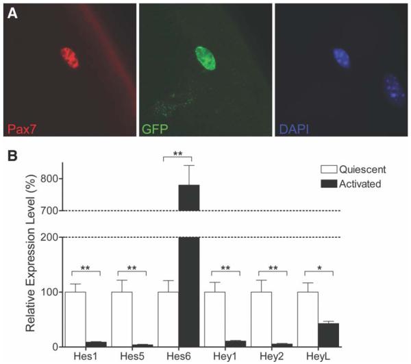
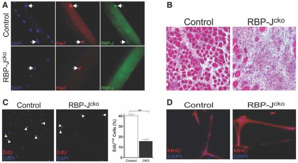
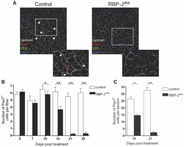
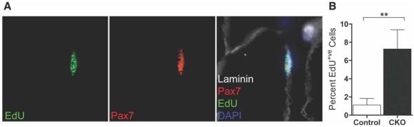
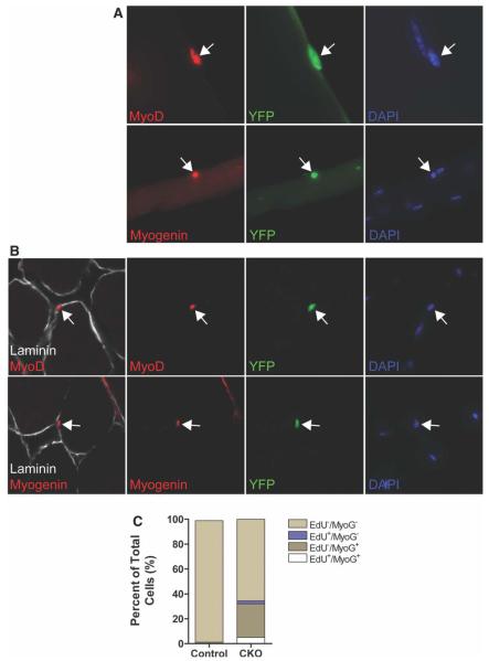
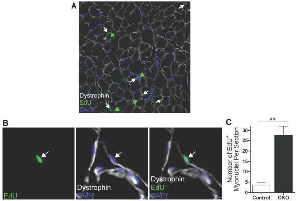
Satellite cells (SCs) are myogenic stem cells found in skeletal muscle that function to repair tissue damaged by injury or disease. SCs are quiescent at rest, although the signaling pathways required to maintain quiescence are unknown. Using a transgenic Notch reporter mouse and quantitative reverse-transcription polymerase chain reaction analysis of Notch target genes, we determined that Notch signaling is active in quiescent SCs. SC-specific deletion of recombining binding protein-Jκ (RBP-Jκ), a nuclear factor required for Notch signaling, resulted in the depletion of the SC pool and muscles that lacked any ability to regenerate in response to injury. SC depletion was not due to apoptosis. Rather, RBP-Jκ-deficient SCs spontaneously activate, fail to self-renew, and undergo terminal differentiation. Intriguingly, most of the cells differentiate without first dividing. They then fuse with adjacent myofibers, leading to the gradual disappearance of SCs from the muscle. These results demonstrate the requirement of Notch signaling for the maintenance of the quiescent state and for muscle stem cell homeostasis by the regulation of self-renewal and differentiation, processes that are all critical for normal postnatal myogenesis.


Figures

Figure 1
Figure 1
Notch signaling is active in quiescent satellite cells (SCs). (A): Immunostaining for Pax7 and GFP in SCs associated with freshly isolated myofibers from a transgenic Notch reporter mouse (×63 magnification). GFP expression in Pax7+ve cells indicates active Notch signaling in quiescent SCs. (B): Quantitative RT-PCR analysis of Notch target gene expression in quiescent and activated SCs. Hes1, Hes5, Hey1, Hey2, and HeyL levels were higher in quiescent SCs. Hes6 was more highly expressed after activation. RNA expression levels are normalized to those at quiescence. (*, p < .05, **, p < .01). Abbreviations: DAPI, 4′,6-diamidino-2-phenylindole; GFP, green fluorescent protein; Pax7, paired box protein 7; RT-PCR, reverse-transcription polymerase chain reaction.
Figure 2
Figure 2
Deletion of RBP-J in SCs leads to a failure of regeneration. (A): Satellite cells (SCs) assayed by immunostaining for the presence of RBP-J protein in control and RBP-Jcko mice after tamoxifen treatment. RBP-J protein is eliminated from Pax7+ve SCs (arrows) in RBP-Jcko mice (−63 magnification). (B): After 4 weeks of the initiation of tamoxifen treatment, RBP-Jcko and control mice were injured using BaCl2 injection and muscles were harvested 7 days later. Cryosections were stained with hematoxylin and eosin and reveal a complete lack of regeneration in the RBP-Jcko muscle. (C): Fluorescence activated cell sorting-purified control and RBP-Jcko SCs from tamoxifen-treated animals 9 days after the completion of tamoxifen injections were tested in vitro for proliferation by the incorporation of EdU. The panels on the left show that both populations underwent proliferative amplification. The graph on the right shows that the population of control SCs expanded at a greater rate than that of the RBP-J-deficient SC population. (**, p < .01). (D): Equivalent numbers of control and RBP-J-deficient SCs were plated and induced to differentiate by culture in low serum medium. Both populations differentiated to produce multinucleate myotubes expressing MHC with no obvious difference between the two populations. Abbreviations: CKO, conditional knockout; DAPI, 4′,6-diamidino-2-phenylindole; EdU, 5-ethynyl-2′-deoxyuridine; MHC, myosin heavy chain; Pax7, paired box protein 7; RBP-J, recombining binding protein-J.
Figure 3
Figure 3
Deletion of RPB-J in satellite cells (SCs) leads to their depletion. (A): Laminin, Pax7, and YFP expression in tibialis anterior muscles from control and RBP-Jcko mice 21 days following tamoxifen treatment. Images to the lower right are higher power views of the areas indicated by the dotted rectangles. YFP+ve SCs are absent in RBP-Jcko animals. (B): Average number of Pax7+ve cells per myofiber as assessed in single fiber cultures from control and tamoxifen-treated RBP-Jcko mice as a function of time after tamoxifen administration. (C): Average number of Pax7+ve cells per cryosection in control and tamoxifen-treated RBP-Jcko mice 14 and 21 days following tamoxifen administration. The decline in SC number paralleled exactly that seen in single myofibers preparations (panel B). (*, p < .05, **, p < .01). Abbreviations: DAPI, 4′,6-diamidino-2-phenylindole; Pax7, paired box protein 7; RBP-J, recombining binding protein-J; YFP, yellow fluorescent protein.
Figure 4
Figure 4
Loss of RBP-J in quiescent satellite cells (SCs) induces cell cycle entry. (A): Control and RBP-Jcko animals were treated with tamoxifen for 5 days and EdU for 14 days, both initiated at the same time. Cryosections of tibialis anterior muscles were assessed for evidence of SC proliferation by EdU incorporation. An EdU+ve cell is shown that is both Pax7+ve and beneath the basal lamina as outlined by laminin staining. (B): Control and RBP-Jcko animals were treated with tamoxifen for 5 days and EdU for 14 days as in panel (A). SCs were purified by fluorescence activated cell sorting, plated, and stained to assess EdU incorporation. The percentages of SCs that were EdU+ve are shown. (**, p < .01). Abbreviations: CKO, conditional knockout; DAPI, 4′,6-diamidino-2-phenylindole; EdU, 5-ethynyl-2′-deoxyuridine; Pax7, paired box protein 7; RBP-J, recombining binding protein-J.
Figure 5
Figure 5
Loss of RBP-J induces satellite cell (SC) activation and differentiation. (A): Muscles were harvested from RBP-Jcko and control mice 10 days after tamoxifen treatment, and myofiber cultures were analyzed for the expression of myogenic activation (by the expression of MyoD) and differentiation (by the expression of Myogenin) in SCs or their progeny identified by YFP expression. Arrows indicate YFP+ve cells that are positive for either MyoD (above) or Myogenin (below). (B): Cryosections from muscles of mice treated as in panel (A), also analyzed for the same markers of activation and differentiation. Arrows indicate YFP+ve cells that express either of the myogenic lineage markers. (C): Fluorescence activated cell sorting-purified SCs from tamoxifen-treated control and RBP-Jcko mice administered EdU for 14 days were plated and immediately assayed for the incorporation of EdU and the expression of Myogenin. In the RBP-J-deficient SC population, more than 25% of the cells were Myogenin+ve, and of those, the vast majority did not incorporate EdU. Abbreviations: CKO, conditional knockout; DAPI, 4′,6-diamidino-2-phenylindole; EdU, 5-ethynyl-2′-deoxyuridine; RBP-J, recombining binding protein-J; YFP, yellow fluorescent protein.
Figure 6
Figure 6
Incorporation of RBP-J-deficient satellite cells (SCs) into adjacent myofibers. (A): Tibialis anterior (TA) muscle cryosection from a RBP-Jcko animal treated daily with EdU for 14 days, beginning at the onset of tamoxifen administration. EdU-labeled myonuclei located beneath the myofiber membrane as delineated by dystrophin staining are highlighted by white arrows. EdU+ve nuclei that are outside the muscle fiber membrane are highlighted by green arrows. (B): Higher power magnification showing a single EdU+ve myonucleus in a cryosection as in panel (A). (C): Quantification of EdU+ve myonuclei, expressed as the absolute number per TA cryosections, from control and RBP-Jcko mice treated as described for panel (A). (**, p < .01). Abbreviations: CKO, conditional knockout; DAPI, 4′,6-diamidino-2-phenylindole; EdU, 5-ethynyl-2′-deoxyuridine; RBP-J, recombining binding protein-J.

References

    1. Charge SB, Rudnicki MA. Cellular and molecular regulation of muscle regeneration. Physiol Rev. 2004;84:209–238. - PubMed
    1. Orford KW, Scadden DT. Deconstructing stem cell self-renewal: Genetic insights into cell-cycle regulation. Nat Rev Genet. 2008;9:115–128. - PubMed
    1. Yusuf I, Fruman DA. Regulation of quiescence in lymphocytes. Trends Immunol. 2003;24:380–386. - PubMed
    1. Coller HA, Sang L, Roberts JM. A new description of cellular quiescence. PLoS Biol. 2006;4:e83. - PMC - PubMed
    1. Chen C, Liu Y, Liu R, et al. TSC-mTOR maintains quiescence and function of hematopoietic stem cells by repressing mitochondrial biogenesis and reactive oxygen species. J Exp Med. 2008;205:2397–2408. - PMC - PubMed

Publication types

MeSH terms

Substances