. 2011 Nov 1;124(Pt 21):3619-30.
doi: 10.1242/jcs.085159. Epub 2011 Nov 1.

Integrity of the network sarcoplasmic reticulum in skeletal muscle requires small ankyrin 1

Affiliations

Integrity of the network sarcoplasmic reticulum in skeletal muscle requires small ankyrin 1

Maegen A Ackermann et al. J Cell Sci. .

Abstract

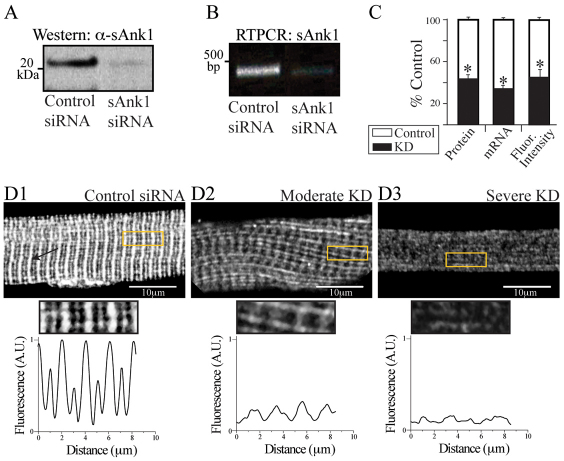
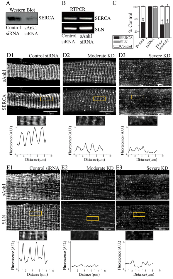
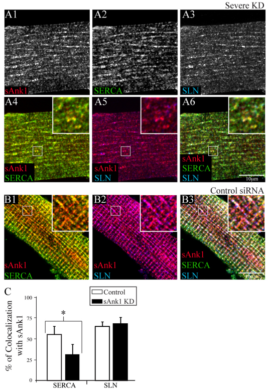
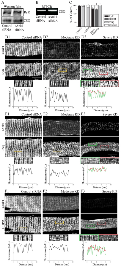
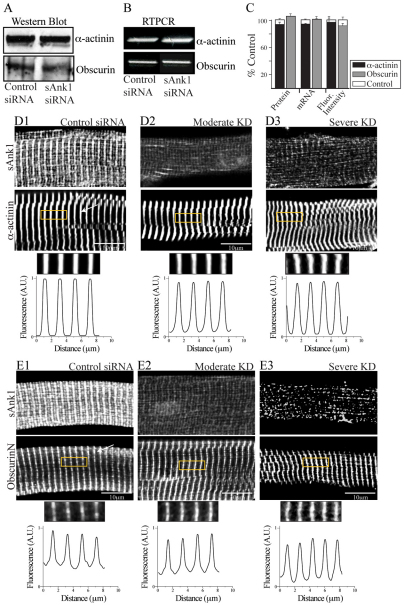
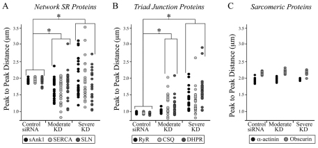
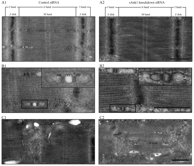
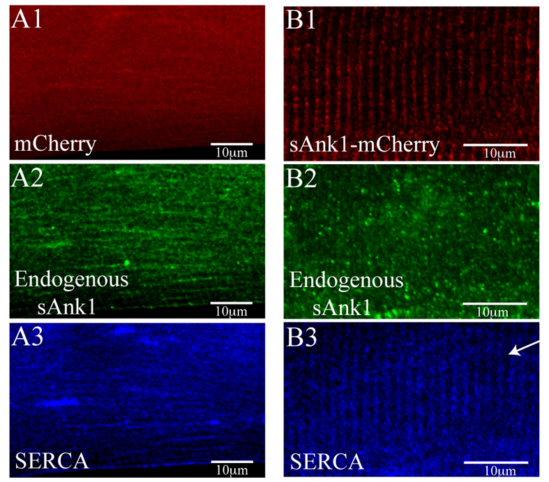
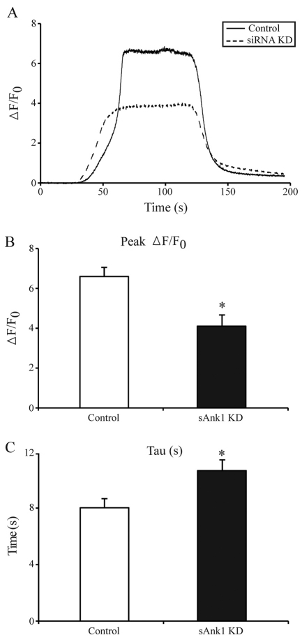
Small ankyrin 1 (sAnk1; Ank1.5) is a ~20 kDa protein of striated muscle that concentrates in the network compartment of the sarcoplasmic reticulum (nSR). We used siRNA targeted to sAnk1 to assess its role in organizing the sarcoplasmic reticulum (SR) of skeletal myofibers in vitro. siRNA reduced sAnk1 mRNA and protein levels and disrupted the organization of the remaining sAnk1. Sarcomeric proteins were unchanged, but two other proteins of the nSR, SERCA and sarcolipin, decreased significantly in amount and segregated into distinct structures containing sarcolipin and sAnk1, and SERCA, respectively. Exogenous sAnk1 restored SERCA to its normal distribution. Ryanodine receptors and calsequestrin in the junctional SR, and L-type Ca(2+) channels in the transverse tubules were not reduced, although their striated organization was mildly altered. Consistent with the loss of SERCA, uptake and release of Ca(2+) were significantly inhibited. Our results show that sAnk1 stabilizes the nSR and that its absence causes the nSR to fragment into distinct membrane compartments.


Figures

Fig. 1.
Fig. 1.
Effect of targeted siRNA on sAnk1 mRNA and protein levels. FDB fibers were transduced with adenovirus encoding siRNA selective for sAnk1 or a control sequence. Samples were maintained in culture for 48 hours and then examined by western blotting, RT-PCR and immunofluorescence. (A,B) sAnk1 protein (A) and mRNA (B) are reduced in cultures of fibers treated with sAnk1-siRNA. (C) Mean sAnk1 protein, cDNA and fluorescent intensity levels are significantly reduced (>50%) in cells treated with sAnk1-siRNA (KD), compared with controls (*P<0.01, Student's t-test). Results are means ± s.e.m. (D) In con-siRNA-treated myofibers (D1), sAnk1 concentrates around Z-disks, and to a lesser extent around M-bands, as indicated by the regular fluorescent peaks. Myofibers treated with sAnk1-siRNA (D2, D3), show a range of intensities and distribution, from little (similar to controls; not shown), to moderate (D2) and severe (D3); 5× regions of interest (ROIs) boxed in yellow. Severely affected fibers (D3) constitute ~15% of the total. Most fibers are only moderately affected (D2), and show reduced labeling for sAnk1, that is absent around the M-band, and present in some longitudinal structures. The arrow in D1 indicates Z-disks. See also supplementary material Fig. S1. The results show that sAnk1-siRNA reduces sAnk1 levels and alters its distribution in skeletal myofibers.
Fig. 2.
Fig. 2.
Reduced expression of sAnk1 leads to disruption of the network SR. FDB myofibers were treated as in Fig. 1 and analyzed for two components of the nSR, SERCA and SLN. (A) SERCA protein is significantly reduced in immunoblots of myofibers treated with sAnk1-siRNA, compared with con-siRNA. (B) Levels of mRNA encoding SERCA and SLN are not altered in myofibers in which sAnk1 expression is reduced. (C) Intensity of immunofluorescence labeling of SERCA and SLN are both significantly reduced (>50%) in cells treated with sAnk1-siRNA, compared with controls. Results are means ± s.e.m. (D,E) The organization of SERCA (D2,D3) and SLN (E2,E3), is disrupted or lost when sAnk1 expression is inhibited as compared with controls (D1 and E1, respectively). Fluorescent profiles of the ROI (yellow boxes) illustrate the normal, striated pattern of SERCA and SLN, which localize primarily at the Z-disks in control fibers (bottom left panels of D1 and E1, respectively), which is lost in fibers with very low levels of sAnk1 (bottom right panels of D3 and E3, respectively). The results show that sAnk1-siRNA reduces the amount of SERCA and SLN proteins, but not their mRNAs, and alters their distribution in skeletal myofibers.
Fig. 3.
Fig. 3.
Residual proteins of the nSR appear in punctate or longitudinal structures when sAnk1 is severely reduced. Myofibers treated as in Fig. 1 were triple labeled for immunofluorescence of sAnk1, SERCA and SLN. (A) Fibers with severely reduced expression of sAnk1 show remaining sAnk1 (A1), SERCA (A2) and SLN (A3) in punctate or longitudinal structures. SERCA and SLN are compared with sAnk1 in color overlays (A4, sAnk1 and SERCA in yellow; A5, sAnk1 and SLN in purple; A6, all three, in white). (B) In control fibers, all proteins colocalize at the Z-disk (sAnk1, red; SERCA, green; and SLN, blue; other colors are as above). (C) Colocalization of each protein with sAnk1 was determined by Pearson's correlation coefficient. In fibers treated with sAnk1-siRNA, the colocalization of SERCA and sAnk1 is significantly reduced (*P<0.01), but colocalization of SLN and sAnk1 is unchanged. The results show that the components of the nSR segregate into distinct structures when sAnk1 is reduced. Data are means ± s.e.m.
Fig. 4.
Fig. 4.
Proteins of the triad junction are mildly altered when sAnk1 is severely reduced. Myofibers were treated as in Fig. 1. Extracts were prepared and analyzed for protein and mRNA levels, or for the distribution of proteins of the jSR (RyR; DHPR; calsequestrin, CSQ). (A) RyR and CSQ polypeptides are unaffected by sAnk1-siRNA. (B) mRNA encoding CSQ is unchanged. (C) Mean protein and mRNA expression and fluorescent intensity levels do not significantly change when sAnk1 is reduced (P<0.01, Student's t-test). Results are means ± s.e.m. (DF) Fluorescent profiles of the boxed ROIs show the normal doublet pattern for RyR, CSQ and DHPR at the A–I junction of control myofibers (bottom panels D1, E1 and F1, respectively) in control fibers. When sAnk1 is moderately reduced, this doublet is disrupted in some regions but stable in most. When sAnk1 is severely reduced, all three antibodies continue to label doublets over many areas of the myoplasm (ROIs in green), but other areas are partially disrupted (ROIs in red). The results show that proteins of the triad junction undergo a small to moderate change in organization when sAnk1 expression is inhibited.
Fig. 5.
Fig. 5.
Sarcomeric proteins are unaffected when sAnk1 is reduced. Myofibers were treated as in Fig. 1 and examined for expression of α-actinin and obscurin. (A) Immunoblot of α-actinin and obscurin indicate that these proteins are unchanged when sAnk1 expression is reduced. (B) RT-PCR shows that the mRNAs encoding α-actinin and obscurin are also unaffected. (C) Quantitative comparisons of the amounts of α-actinin and obscurin and their mRNAs, as well as the intensity of immunofluorescence labeling, reveal no significant differences between controls and myofibers treated to reduce expression of sAnk1 (P<0.01 Student's t-test). Results are means ± s.e.m. (D,E) Immunofluorescence labeling for α-actinin (D) and obscurin (E) reveals no significant differences in the organization of these two proteins in myofibers with either moderately or severely reduced expression of sAnk1. The antibody against obscurin recognizes the N-terminal Ig domains and labels only M-bands. α-Actinin is limited to Z-disks. The results show that the contractile apparatus is not significantly affected by sAnk1-siRNA.
Fig. 6.
Fig. 6.
Quantification of loss of organization of proteins in myofibers with reduced levels of sAnk1. The distribution of proteins in control and siRNA-treated fibers was measured by calculating the average peak-to-peak distances (see Materials and Methods, and supplementary material Fig. S2) and comparing the values seen in control myofibers with those seen in myofibers showing moderate or severe reduction in expression of sAnk1 (n=25 per group). (A) The loss of organization of three proteins of the nSR, sAnk1 (black), SERCA (light grey) and SLN (dark grey) are very similar and differ significantly from controls in populations in which sAnk1 is either moderately or severely reduced (*P<0.01, F-test). (B) Proteins of the jSR, including RyR (black), CSQ (light grey) and DHPR (dark gray) show significant peak-to-peak variation in the myofibers having severely reduced levels of sAnk, compared with controls (*P<0.01, F-test). DHPR and CSQ also show significant variation in myofibers with moderate reduction in sAnk1 (*P<0.01, F-test). (C) Two proteins associated with the contractile apparatus, α-actinin (black) and obscurin (grey) show no significant variation in the average peak-to-peak distances between control and affected groups (P>0.05, F-test).
Fig. 7.
Fig. 7.
Ultrastructure of fibers with reduced levels of sAnk1. FDB myofibers were treated as above, then fixed and processed for thin-section electron microscopy. Views are of the sarcomere (A), triad junctions (B) (comprised of t-tubules flanked on either side by jSR) and nSR (C). Sections through a myofiber treated with con-siRNA show typical structures (A1,B1,C1). Fibers treated with sAnk1-siRNA have sarcomeres and triad junctions that appear normal, as in controls (A2,B2,C2). However, the nSR does not appear as an extensive system of anastomosing tubules overlying the Z-disk, as it does in controls. Instead, it appears reduced in size and complexity, with prominent and enlarged tubular elements organized parallel to the long axis of the fiber (asterisks). Scale bar: 0.5 μm.
Fig. 8.
Fig. 8.
sAnk1–mCherry restores SR structure lost by knockdown of endogenous sAnk1. FDB fibers were transfected in vivo by electroporation, either with a plasmid encoding mCherry (A) or mCherry–sAnk1 (B). One week later, fibers were cultured, treated with sAnk1-siRNA and labeled for immunofluorescence of sAnk1 and SERCA. The results show that, although the knockdown of expression of endogenous sAnk1 in these fibers was considerable and resulted in significant disorganization of the remaining sAnk1 (A2 and B2), the striated organization of SERCA in the nSR is specifically rescued by sAnk1-mCherry (B3) but not by mCherry alone (A3). Scale bar: 10 μm. Arrow indicates Z-disk.
Fig. 9.
Fig. 9.
Ca2+ transients in fibers with reduced levels of sAnk1. FDB fibers treated as in Fig. 1 were loaded with Fluo-4 to measure the [Ca2+]i transient induced by exposure to the RyR1 agonist, 4-CmC (1 mM). (A) Representative fluorescence profiles are of confocal line-scan images from con-siRNA-treated (solid line) and sAnk1-siRNA-treated (dashed line) myofibers. The latter shows lower peak fluorescence. (B) Data from three experiments, performed as in A were pooled. The peak of the [Ca2+]i transient in fibers treated with sAnk1-siRNA is significantly smaller than controls (*P<0.01). (C) Following washout of 4-CmC, the rapid decay phase of the [Ca2+]i transient was fitted by a single-exponential to determine tau. Tau of the [Ca2+]i transient is significantly slower in sAnk1-siRNA-treated myofibers than in controls (*P<0.01). The data show that the size and rate of refilling of Ca2+ stores in FDB myofibers are reduced when sAnk1 expression is inhibited by siRNA. Results are means ± s.e.m.

References

    1. Ackermann M. A., Hu L. Y., Bowman A. L., Bloch R. J., Kontrogianni-Konstantopoulos A. (2009). Obscurin interacts with a novel isoform of MyBP-C slow at the periphery of the sarcomeric M-band and regulates thick filament assembly. Mol. Biol. Cell 20, 2963-2978 - PMC - PubMed
    1. Babu G. J., Bhupathy P., Timofeyev V., Petrashevskaya N. N., Reiser P. J., Chiamvimonvat N., Periasamy M. (2007). Ablation of sarcolipin enhances sarcoplasmic reticulum calcium transport and atrial contractility. Proc. Natl. Acad. Sci. USA 104, 17867-17872 - PMC - PubMed
    1. Bagnato P., Barone V., Giacomello E., Rossi D., Sorrentino V. (2003). Binding of an ankyrin-1 isoform to obscurin suggests a molecular link between the sarcoplasmic reticulum and myofibrils in striated muscles. J. Cell Biol. 160, 245-253 - PMC - PubMed
    1. Birkenmeier C. S., Sharp J. J., Gifford E. J., Deveau S. A., Barker J. E. (1998). An alternative first exon in the distal end of the erythroid ankyrin gene leads to production of a small isoform containing an NH2-terminal membrane anchor. Genomics 50, 79-88 - PubMed
    1. Borzok M. A., Catino D. H., Nicholson J. D., Kontrogianni-Konstantopoulos A., Bloch R. J. (2007). Mapping the binding site on small ankyrin 1 for obscurin. J. Biol. Chem. 282, 32384-32396 - PubMed

Publication types

MeSH terms