Primary vitreoretinal lymphoma: a report from an International Primary Central Nervous System Lymphoma Collaborative Group symposium
- PMID: 22045784
- PMCID: PMC3233294
- DOI: 10.1634/theoncologist.2011-0210
Primary vitreoretinal lymphoma: a report from an International Primary Central Nervous System Lymphoma Collaborative Group symposium
Abstract
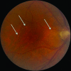
Primary vitreoretinal lymphoma (PVRL), also known as primary intraocular lymphoma, is a rare malignancy typically classified as a diffuse large B-cell lymphoma and most frequently develops in elderly populations. PVRL commonly masquerades as posterior uveitis and has a unique tropism for the retina and central nervous system (CNS). Over 15% of primary CNS lymphoma patients develop intraocular lymphoma, usually occurring in the retina and/or vitreous. Conversely, 65%-90% of PVRL patients develop CNS lymphoma. Consequently, PVRL is often fatal because of ultimate CNS association. Current PVRL animal models are limited and require further development. Typical clinical findings include vitreous cellular infiltration (lymphoma and inflammatory cells) and subretinal tumor infiltration as determined using dilated fundoscopy, fluorescent angiography, and optical coherent tomography. Currently, PVRL is most often diagnosed using both histology to identify lymphoma cells in the vitreous or retina and immunohistochemistry to indicate monoclonality. Additional adjuncts in diagnosing PVRL exist, including elevation of interleukin-10 levels in ocular fluids and detection of Ig(H) or T-cell receptor gene rearrangements in malignant cells. The optimal therapy for PVRL is not defined and requires the combined effort of oncologists and ophthalmologists. PVRL is sensitive to radiation therapy and exhibits high responsiveness to intravitreal methotrexate or rituximab. Although systemic chemotherapy alone can result in high response rates in patients with PVRL, there is a high relapse rate. Because of the disease rarity, international, multicenter, collaborative efforts are required to better understand the biology and pathogenesis of PVRL as well as to define both diagnostic markers and optimal therapies.
Conflict of interest statement
Figures


References
-
- Chan CC, Gonzalez JA. Primary Intraocular Lymphoma. New Jersey, London, Singapore, Beijing, Shanghai, Hong Kong, Taipei: Chennai World Scientific Publishing Co. Pte. Ltd.; 2007. pp. 1–267.
-
- Coupland SE, Damato B. Understanding intraocular lymphomas. Clin Experiment Ophthalmol. 2008;36:564–578. - PubMed
-
- Cummings TJ, Stenzel TT, Klintworth G, et al. Primary intraocular T-cell-rich large B-cell lymphoma. Arch Pathol Lab Med. 2005;129:1050–1053. - PubMed
Publication types
MeSH terms
Grants and funding
LinkOut - more resources
Full Text Sources

