. 2011 Dec 30;286(52):44295-305.
doi: 10.1074/jbc.M111.258053. Epub 2011 Nov 1.

Mammalian target of rapamycin complex 2 (mTORC2) negatively regulates Toll-like receptor 4-mediated inflammatory response via FoxO1

Affiliations

Mammalian target of rapamycin complex 2 (mTORC2) negatively regulates Toll-like receptor 4-mediated inflammatory response via FoxO1

Jonathan Brown et al. J Biol Chem. .

Abstract

Activation of the PI3K pathway plays a pivotal role in regulating the inflammatory response. The loss of mTORC2 has been shown to abrogate the activation of Akt, a critical downstream component of PI3K signaling. However, the biological importance of mTORC2 in innate immunity is currently unknown. Here we demonstrate that rictor, a key component of mTORC2, plays a critical role in controlling the innate inflammatory response via its ability to regulate FoxO1. Upon LPS stimulation, both rictor-deficient mouse embryonic fibroblasts (MEFs) and rictor knockdown dendritic cells exhibited a hyperinflammatory phenotype. The hyperinflammatory phenotype was due to a defective Akt signaling axis, because both rictor-deficient MEFs and rictor knockdown dendritic cells exhibited attenuated Akt phosphorylation and kinase activity. Analysis of downstream Akt targets revealed that phosphorylation of FoxO1 was impaired in rictor-deficient cells, resulting in elevated nuclear FoxO1 levels and diminished nuclear export of FoxO1 upon LPS stimulation. Knockdown of FoxO1 attenuated the hyperinflammatory phenotype exhibited by rictor-deficient MEFs. Moreover, FoxO1 deletion in dendritic cells attenuated the capacity of LPS to induce inflammatory cytokine expression. These findings identify a novel signaling pathway by which mTORC2 regulates the TLR-mediated inflammatory response through its ability to regulate FoxO1.


Figures

FIGURE 1.
FIGURE 1.
Genetic loss or siRNA knockdown of rictor results in defective Akt phosphorylation and activity. A and B, wild type and rictor-deficient MEFs were stimulated with LPS over a 2-h time course, and levels of rictor, raptor, and mLST8 (A) and total mTOR (B) were monitored by immunoblot. C and D, phosphorylation levels of Akt Ser-473 in wild type and rictor-deficient MEFs stimulated with LPS were monitored by immunoblot (C) and ELISA (D) using whole cell lysates. E, dendritic cells were plated at a concentration of 1 × 10⁶/ml, rested overnight, and then treated with control or rictor siRNA for 3 days. The cellular levels of rictor were assessed by immunoblot 3 days post-transfection. F, the phosphorylation levels of Akt Ser-473 were assessed by ELISA in LPS-stimulated DC treated with control or rictor siRNA. G, wild type and rictor-deficient MEFs were stimulated with LPS for up to 60 min and lysed, total Akt was immunoprecipitated, and immunoprecipitated Akt was incubated with a GSK3 fusion protein as substrate. Akt activity, as determined by phospho-GSK3 α/β (Ser-9) levels, was assessed by immunoblot. For the Akt activity assay, all of the immunoblots were normalized to total pull-down of Akt. The levels of β-actin were monitored to ensure equivalent sample loading. For A–C, E, and G, the data are representative of three to five separate experiments. For D and F, the data represent the arithmetic means ± S.D. of three separate experiments. *** indicates statistical significance at p < 0.001.
FIGURE 2.
FIGURE 2.
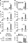
Rictor-deficient MEFs or DC treated with rictor siRNA exhibit a hyperinflammatory response when stimulated with LPS. A and B, rictor-deficient MEFs stimulated with LPS displayed elevated levels of IL-6 (A) and IL-8 production (B), as compared with wild type control MEFs. C–F, siRNA knockdown of rictor in DC leads to increased levels of IL-1β (C), TNF-α (D), IL-6 (E), and IL-12 p70 (F) upon LPS stimulation, as compared with control siRNA dendritic cells stimulated with LPS. G, transfection of Myr-Akt into rictor-deficient MEFs resulted in elevated levels of Akt, as compared with rictor-deficient MEFs transfected with a control plasmid. β-Actin was monitored to ensure equivalent sample loading. H, transfection of Myr-Akt into rictor-deficient MEFs attenuated the enhanced levels of IL-6 production when stimulated with LPS, as compared with rictor-deficient MEFs transfected with a control plasmid. Transfection of rictor-deficient MEFs with a control plasmid did not significantly affect the LPS-induced production of IL-6, as compared with nontransfected rictor-deficient MEFs stimulated with LPS. The data are representative of three to five separate experiments. For A–F and H, the data represent the arithmetic mean ± S.D. of five separate experiments. For G, the data are representative of three separate experiments. ** and *** indicate statistical significance at p < 0.01 and p < 0.001, respectively. Ctrl, control.
FIGURE 3.
FIGURE 3.
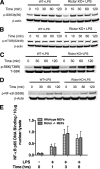
Rictor is not required for the phosphorylation of GSK3, mTOR, p70S6K, or NF-κB in LPS-stimulated cells. A–D, the levels of phospho-GSK3-β (A), mTOR (B), p70S6K (C), and NF-κB (D) p65 were determined by immunoblot in wild type and rictor-deficient MEFs stimulated with LPS. E, wild type and rictor-deficient MEFs were stimulated with LPS for 6 h, and nuclear lysates were assessed for NF-κB p65 DNA binding. The data are representative of three to five separate experiments.
FIGURE 4.
FIGURE 4.
Rictor is required for the phosphorylation of PKC-α and SGK but does not account for the hyperinflammatory phenotype observed in rictor-deficient cells simulated with LPS. A and B, the levels of phospho-SGK-1 (A) and phospho-PKC-α (B) were determined by immunoblot of whole cell lysates isolated from LPS-stimulated wild type and rictor-deficient MEFs. C and D, wild type and rictor-deficient MEFs were pretreated with an SGK inhibitor (C) or PKC-α inhibitor (D) for 1 h and stimulated with LPS, and cell-free supernatants were monitored for IL-6 levels by ELISA. For A and B, the data are representative of three separate experiments. In C and D, the data represent the arithmetic means ± S.D. of five separate experiments. ** indicates statistical significance at p < 0.01.
FIGURE 5.
FIGURE 5.
FoxO1 phosphorylation, nuclear localization, and transcriptional activity are defective in rictor-deficient cells. A, FoxO1 phosphorylation levels in wild type and rictor-deficient MEFs stimulated with LPS. B, siRNA knockdown of rictor in LPS-stimulated DC resulted in defective FoxO1 phosphorylation, as compared with control siRNA-treated DC stimulated with LPS. C, after siRNA-mediated knockdown of rictor, dendritic cells were stimulated with LPS for 6 h, and nuclear lysates were monitored for FoxO1 levels by immunoblot. D, wild type and rictor-deficient MEFs were stimulated with LPS for 6 h time, and nuclear lysates were monitored for FoxO1 DNA binding activity. E, rictor-deficient and wild type MEFs were transfected with a luciferase reporter plasmid for FoxO1 and a control plasmid expressing β-galactosidase. The cells were stimulated with LPS for 20 h, and luciferase activity was assessed and reported as the ratio of luciferase to β-galactosidase. For A–C, the data are representative of three separate experiments. For D and E, the data represent the arithmetic mean ± S.D. of five separate experiments. ** and *** indicate statistical significance at p < 0.01 and p < 0.001, respectively.
FIGURE 6.
FIGURE 6.
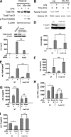
Complementation with constitutively active Akt or knockdown of FoxO1 abrogates the hyperinflammatory response of LPS-stimulated rictor-deficient cells. A and B, LPS-stimulated, rictor-deficient MEFs have restored FoxO1 phosphorylation (Ser-256) (A) and nuclear export of FoxO1 (B) when transfected with Myr-Akt compared with rictor-deficient MEFs transfected with a control plasmid and LPS. C, siRNA was utilized to knockdown FoxO1 in rictor-deficient MEFs. Knockdown of FoxO1 by siRNA in rictor-deficient MEFs resulted in decreased production of IL-6 upon LPS stimulation compared with LPS-stimulated, rictor-deficient MEFs treated with control siRNA as assessed by ELISA. D–F, WT and FoxO1 KO DC were stimulated with LPS for 20 h, and IL-6 (D), IL-12p40 (E), and IL-10 (F) levels were assessed by ELISA. G–I, rictor was knocked down by siRNA in both WT and FoxO1 KO DC. Three days after transfection, the DC were stimulated with LPS for 20 h. IL-6 (G), TNF-α (H), and IL-10 (I) levels were determined by ELISA. For A–I, the data are representative of three to five separate experiments. For C–I, the data represent the arithmetic means ± S.D. of five separate experiments. ** and *** indicate statistical significance at p < 0.01 and p < 0.001, respectively. Ctrl, control; NS, not significant.

References

    1. Cantley L. C. (2002) Science 296, 1655–1657 - PubMed
    1. Alessi D. R., James S. R., Downes C. P., Holmes A. B., Gaffney P. R., Reese C. B., Cohen P. (1997) Curr. Biol. 7, 261–269 - PubMed
    1. Stephens L., Anderson K., Stokoe D., Erdjument-Bromage H., Painter G. F., Holmes A. B., Gaffney P. R., Reese C. B., McCormick F., Tempst P., Coadwell J., Hawkins P. T. (1998) Science 279, 710–714 - PubMed
    1. Persad S., Attwell S., Gray V., Mawji N., Deng J. T., Leung D., Yan J., Sanghera J., Walsh M. P., Dedhar S. (2001) J. Biol. Chem. 276, 27462–27469 - PubMed
    1. Feng J., Park J., Cron P., Hess D., Hemmings B. A. (2004) J. Biol. Chem. 279, 41189–41196 - PubMed

Publication types

MeSH terms