. 2011 Dec 30;286(52):45000-13.
doi: 10.1074/jbc.M111.270934. Epub 2011 Nov 1.

Protein-tyrosine phosphatase 1B modulates early endosome fusion and trafficking of Met and epidermal growth factor receptors

Affiliations

Protein-tyrosine phosphatase 1B modulates early endosome fusion and trafficking of Met and epidermal growth factor receptors

Veena Sangwan et al. J Biol Chem. .

Abstract

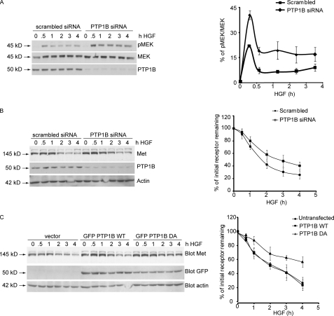
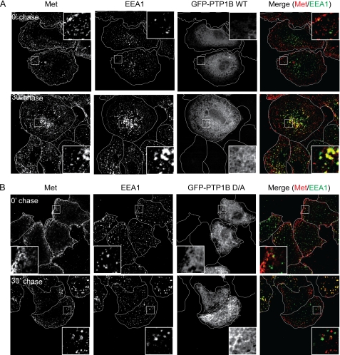
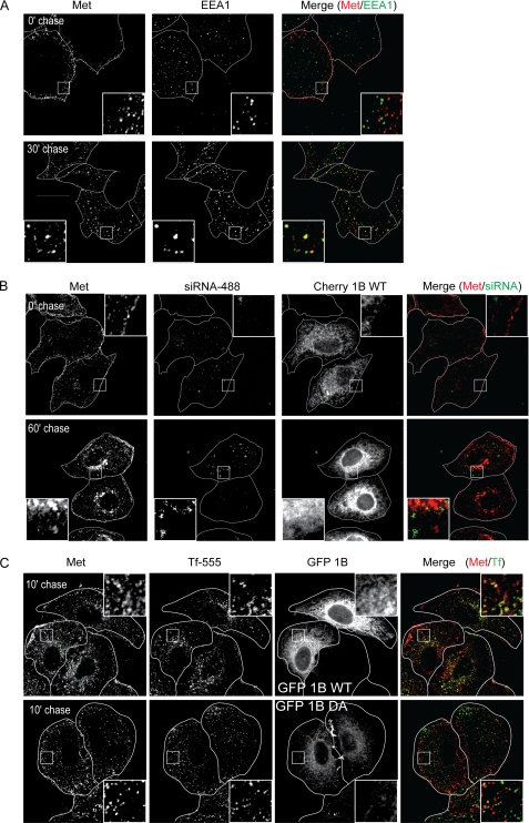
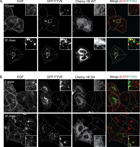
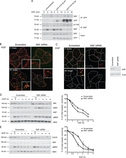
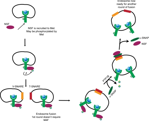
The endoplasmic reticulum-localized non-receptor protein-tyrosine phosphatase 1B (PTP1B) is associated with oncogenic, metabolic, and cytokine-related signaling and functionally targets multiple receptor tyrosine kinases (RTKs) for dephosphorylation. Loss of PTP1B activity leads to enhanced ligand-dependent biological activity of the Met RTK among others. Here, we demonstrate that knockdown of PTP1B or expression of a PTP1B trapping aspartic acid-to-alanine substitution (D/A) mutant delayed ligand-induced degradation of the Met and EGF RTKs. Loss of PTP1B function abrogated trafficking of Met and EGF receptor to Rab5- and phosphatidylinositol 3-phosphate (Pl3P)-positive early endosomes and subsequent trafficking through the degradative pathway. Under these conditions, internalization of the Met and EGF receptors was unaltered, suggesting a block at the level of early endosome formation. We show that the N-ethylmaleimide-sensitive factor (NSF), an essential component of the vesicle fusion machinery, was hyperphosphorylated in PTP1B knockdown or PTP1B D/A-expressing cells and was a target for PTP1B. NSF knockdown phenocopied PTP1B knockdown, demonstrating a mechanism through which PTP1B regulates endocytic trafficking. Finally, we show that PTP1B dephosphorylated NSF and that this interaction was required for physiological RTK trafficking and appropriate attenuation of downstream signaling.


Figures

FIGURE 1.
FIGURE 1.
Loss of function of PTP1B leads to protracted Met signaling and delayed Met dephosphorylation and degradation following HGF stimulation but does not affect Met ubiquitination. A, HeLa cells were transfected either with scrambled siRNA or siRNA against PTP1B and stimulated with HGF for the times indicated. Protein lysates from these cells were subjected to immunoblotting with antibody against MEK, pMEK, or PTP1B. The graph on the right depicts the percentage of phosphorylated MEK. B, HeLa cells were transfected either with scrambled siRNA or siRNA against PTP1B and stimulated with HGF for the times indicated. Protein lysates from these cells were subjected to immunoblotting with antibody against Met, PTP1B, or actin (loading control). The graph on the right depicts the percentage of initial receptor remaining. C, HeLa cells were transfected with either GFP-PTP1B WT or the mutant GFP-PTP1B D/A and stimulated with HGF in the presence of cycloheximide for the times indicated. Protein lysates from these cells were subjected to immunoblotting with antibody against Met, GFP, or actin (loading control). The graph on the right depicts the percentage of initial receptor remaining.
FIGURE 2.
FIGURE 2.
Loss of function of PTP1B delays Met trafficking following HGF stimulation. A and B, HeLa cells were seeded on glass coverslips and transfected with either GFP-PTP1B WT (A) or D/A (B) 16 h postseeding. 24 h later, cells were serum-starved in the presence of cycloheximide for 2 h and loaded with HGF at 4 °C for 1 h. Cells were chased with warm medium without HGF and fixed at the times indicated followed by staining with anti-Met and anti-EEA1 antibodies. The merge of Met (red) and EEA1 (green) is shown in GFP-expressing cells. Pearson's coefficient of co-localization, 0.17 ± 0.01 (0 min (0′)) and 0.61 ± 0.02 (30 min (30′)) for PTP1B WT-transfected cells and 0.13 ± 0.03 (0 min) and 0.16 ± 0.03 (30 min) for PTP1B D/A-transfected cells. p values, 0.2550 (0 min) and 0.0002 (30 min).
FIGURE 3.
FIGURE 3.
Loss of PTP1B delays Met trafficking but does not affect transferrin recycling following HGF stimulation. HeLa cells were seeded on coverslips and transfected with siRNA directed against PTP1B (A). siRNA-treated cells were rescued by overexpression of cherry-PTP1B (Cherry 1B) WT 24 h post-siRNA transfection (B). 48 h post-siRNA transfection, cells were serum-starved in the presence of cycloheximide for 2 h and loaded with HGF at 4 °C for 1 h. Cells were chased with warm medium without HGF and fixed at the times indicated followed by staining with anti-Met and anti-EEA1 antibodies. A, PTP1B siRNA-treated cells. The merge of Met (red) and EEA1 (green) is shown. Pearson's coefficient of co-localization for PTP1B siRNA transfected cells, 0.11 ± 0.01 (0 min (0′)) and 0.46 ± 0.03 (30 min (30′)). p values, 0.0697 (0 min) and 0.7663 (30 min). B, Alexa Fluor 488-labeled PTP1B siRNA-treated cells overexpressing cherry-PTP1B WT. Trafficking of Met to the perinuclear compartment in cells overexpressing siRNA-resistant cherry-PTP1B WT is shown. C, HeLa cells were seeded on coverslips and transfected with GFP-PTP1B WT. Cells were loaded with HGF and Alexa Fluor 555-transferrin (Tf-555), allowed to internalize, and chased for the times indicated. Cells were fixed and stained for extracellular Met. Pearson's coefficient of co-localization, 0.49 ± 0.01 (10 min) for PTP1B WT-transfected cells and 0.28 ± 0.05 (10 min) for PTP1B D/A-transfected cells. p values, 0.5669 (0 min) and 0.1493 (30 min).
FIGURE 4.
FIGURE 4.
Loss of function of PTP1B leads to delayed EGFR degradation and protracted EGFR signaling following EGF stimulation. A, HeLa cells were transfected with either GFP-PTP1B WT or the mutant GFP-PTP1B D/A and stimulated with EGF in the presence of cycloheximide for the times indicated. Protein lysates from these cells were subjected to immunoblotting with antibodies against EGFR, GFP (transfection control), and actin (loading control). B, HeLa cells were seeded on glass coverslips and transfected with either GFP-PTP1B WT or D/A 16 h postseeding. 24 h later, cells were serum-starved in the presence of cycloheximide for 2 h and loaded with Alexa Fluor 555-EGF at 4 °C for 1 h. Cells were chased with warm medium without EGF and fixed at the times indicated followed by staining with an anti-EEA1 antibody. The merge of EGF (red) and EEA1 (green) is shown. Pearson's coefficient of co-localization, 0.13 ± 0.02 (0 min (0′)) and 0.46 ± 0.02 (30 min (30′)) for PTP1B WT-transfected cells and 0.14 ± 0.02 (0 min) and 0.22 ± 0.04 (30 min) for PTP1B D/A-transfected cells. p values, 0.6702 (0 min) and 0.0322 (30 min). C, HeLa cells were seeded on glass coverslips and transfected with either scrambled or PTP1B-specific siRNA 16 h postseeding. 24 h later, cells were serum-starved in the presence of cycloheximide for 2 h and loaded with Alexa Fluor 555-EGF at 4 °C for 1 h. Cells were chased with warm medium without EGF and fixed at the times indicated followed by staining with an anti-EEA1 antibody. The merge of EGF (red) and EEA1 (green) is shown. Pearson's coefficient of co-localization, 0.14 ± 0.02 (0 min) and 0.37 ± 0.03 (30 min) for scrambled siRNA-transfected cells and 0.08 ± 0.04 (0 min) and 0.49 ± 0.03 (30 min) for PTP1B siRNA-transfected cells. p values, 0.2766 (0 min) and 0.0427 (30 min). D, HeLa cells were transfected either with scrambled siRNA or siRNA against PTP1B and stimulated with EGF for the times indicated. Protein lysates from these cells were subjected to immunoblotting with antibodies against pMEK, MEK, PTP1B, and actin (loading control). E, HeLa cells were seeded on coverslips and transfected with GFP-PTP1B WT. Cells were loaded with Alexa Fluor 647-EGF and Alexa Fluor 555-transferrin (Tf-555), allowed to internalize, chased for the times indicated, and fixed. Pearson's coefficient of co-localization, 0.49 ± 0.02 (10 min) for PTP1B WT-transfected cells and 0.35 ± 0.03 (10 min) for PTP1B D/A-transfected cells. p values, 0.8252 (0 min) and 0.0255 (30 min).
FIGURE 5.
FIGURE 5.
Met does not reach FYVE-positive compartment in HeLa cells overexpressing PTP1B D/A. HeLa cells were seeded on coverslips and at 16 h postseeding transfected with cherry-PTP1B (Cherry 1B) WT (A) or D/A (B) and GFP-FYVE. 20 h post-transfection, cells were serum-starved in the presence of cycloheximide for 2 h and loaded with HGF at 4 °C for 1 h. Cells were chased with warm medium without HGF and fixed at the times indicated followed by staining with an antibody directed against the extracellular region of Met. A, GFP-FYVE- and cherry-PTP1B WT-overexpressing cells. The merge of Met (red) and FYVE (green) is shown. B, GFP-FYVE- and cherry-PTP1B D/A-overexpressing cells. The merge of Met (red) and FYVE (green) is shown. Pearson's coefficient of co-localization, 0.07 ± 0.02 (0 min (0′)), 0.47 ± 0.08 (15 min (15′)), and 0.35 ± 0.02 (30 min (30′)) for PTP1B WT-transfected cells and 0.11 ± 0.02 (0 min), 0.18 ± 0.02 (15 min), and 0.16 ± 0.04 (30 min) for PTP1B D/A-transfected cells. p values, 0.2145 (0 min), 0.0259 (15 min), and 0.0232 (30 min).
FIGURE 6.
FIGURE 6.
EGF does not reach FYVE-positive compartment in HeLa cells overexpressing PTP1B D/A. HeLa cells were seeded on coverslips and at 16 h postseeding transfected with cherry-PTP1B (Cherry-1B) WT (A) or D/A (B) and GFP-FYVE. 16 h post-transfection, cells were serum-starved in the presence of cycloheximide for 2 h and loaded with Alexa Fluor 647-EGF at 4 °C for 1 h. Cells were chased with warm medium without EGF. A, GFP-FYVE- and cherry-PTP1B WT-overexpressing cells. The merge of EGF (red) and FYVE (green) is shown. B, GFP-FYVE- and cherry-PTP1B D/A-overexpressing cells. The merge of EGF (red) and FYVE (green) is shown. Pearson's coefficient of co-localization, 0.07 ± 0.02 (0 min (0′)) and 0.44 ± 0.06 (30 min (30′)) for PTP1B WT-transfected cells and 0.05 ± 0.02 (0 min) and 0.18 ± 0.01 (30 min) for PTP1B D/A-transfected cells. p values, 0.4359 (0 min) and 0.0132 (30 min).
FIGURE 7.
FIGURE 7.
NSF is substrate of PTP1B, and knockdown of NSF delays RTK trafficking and protracts signaling. A, immunoblotting of PTP1B immunoprecipitates with the anti-NSF antibody shows that NSF is trapped by PTP1B. HeLa cells were transfected with either GFP-PTP1B WT or GFP-PTP1B D/A and stimulated with HGF in the presence of cycloheximide for the times indicated. Serial immunoprecipitations were performed with anti-GFP and anti-NSF antibodies, and immunoprecipitates and total protein lysates from these cells were subjected to immunoblotting with antibodies against NSF, GFP, and phosphotyrosine Tyr(P)-100 (pY100). Note that NSF is hyperphosphorylated in the absence of PTP1B and is unaffected by presence of the Met ligand, HGF. B, HeLa cells were seeded on coverslips and at 16 h postseeding transfected with either siRNA against NSF or a scrambled siRNA control. 48 h post-transfection, cells were serum-starved in the presence of cycloheximide for 2 h and loaded with HGF at 4 °C for 1 h. Cells were chased with warm medium without HGF and fixed at the times indicated followed by staining with an antibody against the extracellular region of Met. The merge of EEA1 (green) and Met (red) is shown. Side panel, immunoblot depicting knockdown of NSF for this experiment. Pearson's coefficient of co-localization, 0.05 ± 0.02 (0 min (0′)) and 0.45 ± 0.02 (30 min (30′)) for scrambled siRNA-transfected cells and 0.13 ± 0.01 (0 min) and 0.44 ± 0.07 (30 min) for NSF1 siRNA-transfected cells. p values, 0.0330 (0 min) and 0.9188 (30 min). C, HeLa cells were seeded on coverslips and at 16 h postseeding transfected with either siRNA against NSF or a scrambled siRNA control. 48 h post-transfection, cells were serum-starved in the presence of cycloheximide for 2 h and loaded with Alexa Fluor 555-EGF at 4 °C for 1 h. Cells were chased with warm medium without HGF and fixed at the times indicated followed by staining with an antibody against the extracellular region of Met. The merge of EEA1 (green) and Met (red) is shown. Pearson's coefficient of co-localization, 0.09 ± 0.02 (0 min) and 0.51 ± 0.01 (30 min) for scrambled siRNA-transfected cells and 0.12 ± 0.04 (0 min) and 0.52 ± 0.03 (30 min) for NSF1 siRNA-transfected cells. p values, 0.5462 (0 min) and 0.7938 (30 min). D, HeLa cells transfected with either scrambled siRNA or siRNA against PTP1B were serum-starved overnight and stimulated with HGF in the presence of cycloheximide for the times indicated. Protein lysates from these samples were subjected to immunoblotting with antibodies against Met, phosphorylated Met (pMet), NSF, phosphorylated MEK, and total MEK. The graph on the right depicts the percentage of initial Met receptor remaining. E, HeLa cells transfected with either scrambled siRNA or siRNA against NSF were serum-starved overnight and stimulated with EGF in the presence of cycloheximide for the times indicated. Protein lysates from these samples were subjected to immunoblotting with antibodies against EGFR, NSF, phosphorylated MEK, and total MEK. Note that the loss of NSF results in delayed RTK degradation and sustained pMEK activation.
FIGURE 8.
FIGURE 8.
Proposed model for regulation of PTP1B and NSF-mediated vesicle fusion during endocytosis of Met RTK.

References

    1. Sorkin A., von Zastrow M. (2009) Nat. Rev. Mol. Cell Biol. 10, 609–622 - PMC - PubMed
    1. Peschard P., Park M. (2003) Cancer Cell 3, 519–523 - PubMed
    1. Abella J. V., Park M. (2009) Am. J. Physiol. Endocrinol. Metab. 296, E973–E984 - PubMed
    1. Hammond D. E., Urbé S., Vande Woude G. F., Clague M. J. (2001) Oncogene 20, 2761–2770 - PubMed
    1. Hammond D. E., Carter S., McCullough J., Urbé S., Vande Woude G., Clague M. J. (2003) Mol. Biol. Cell 14, 1346–1354 - PMC - PubMed

Publication types

MeSH terms

Substances