. 2011 Dec 23;286(51):44057-44066.
doi: 10.1074/jbc.M111.236711. Epub 2011 Nov 1.

Degradation of p21Cip1 through anaphase-promoting complex/cyclosome and its activator Cdc20 (APC/CCdc20) ubiquitin ligase complex-mediated ubiquitylation is inhibited by cyclin-dependent kinase 2 in cardiomyocytes

Affiliations

Degradation of p21Cip1 through anaphase-promoting complex/cyclosome and its activator Cdc20 (APC/CCdc20) ubiquitin ligase complex-mediated ubiquitylation is inhibited by cyclin-dependent kinase 2 in cardiomyocytes

Kazuhiko Yamada et al. J Biol Chem. .

Abstract

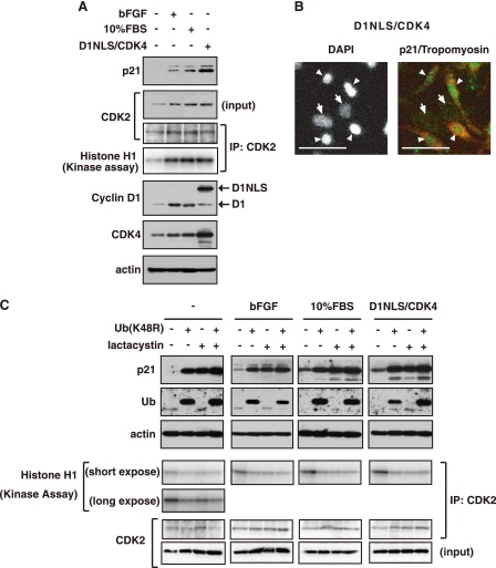
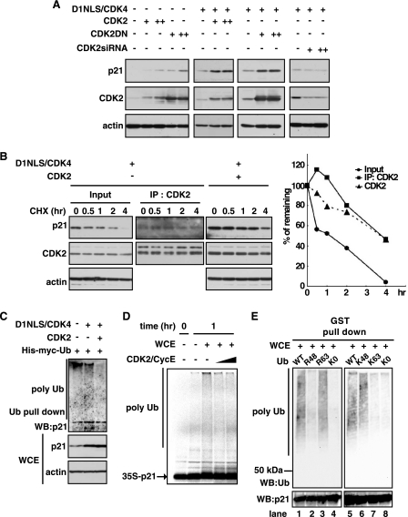
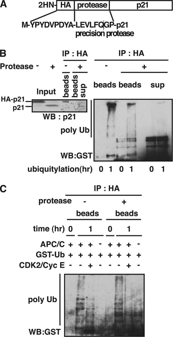
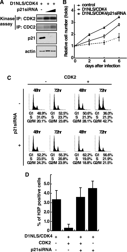
Cyclin-dependent kinase inhibitor p21Cip1 plays a crucial role in regulating cell cycle arrest and differentiation. It is known that p21Cip1 increases during terminal differentiation of cardiomyocytes, but its expression control and biological roles are not fully understood. Here, we show that the p21Cip1 protein is stabilized in cardiomyocytes after mitogenic stimulation, due to its increased CDK2 binding and inhibition of ubiquitylation. The APC/CCdc20 complex is shown to be an E3 ligase mediating ubiquitylation of p21Cip1 at the N terminus. CDK2, but not CDC2, suppressed the interaction of p21Cip1 with Cdc20, thereby leading to inhibition of anaphase-promoting complex/cyclosome and its activator Cdc20 (APC/CCdc20)-mediated p21Cip1 ubiquitylation. It was further demonstrated that p21Cip1 accumulation caused G2 arrest of cardiomyocytes that were forced to re-enter the cell cycle. Taken together, these data show that the stability of the p21Cip1 protein is actively regulated in terminally differentiated cardiomyocytes and plays a role in inhibiting their uncontrolled cell cycle progression. Our study provides a novel insight on the control of p21Cip1 by ubiquitin-mediated degradation and its implication in cell cycle arrest in terminal differentiation.


Figures

FIGURE 1.
FIGURE 1.
p21 is stabilized in cardiomyocytes in response to mitogenic stimuli. Cardiomyocytes were treated with 10 ng of bFGF, 10% FBS for 24 h or infected with a combination of adenoviruses encoding D1NLS and CDK4 for 48 h. A, Western blotting of whole cell extracts for p21, CDK2, cyclin D1, CDK4, sarcomeric actin, and CDK2 in immunoprecipitates (IP) with anti-CDK2 antibody and kinase activity of CDK2. B, immunostaining for p21 in D1NLS/CDK4-expressing cardiomyocytes. Green, p21; red, tropomyosin; white, DAPI. Arrowheads and arrows indicate cardiomyocytes and cardiac fibroblasts, respectively. Scale bar, 50 μm. C, Western blotting of whole cell extracts for p21, ubiquitin, sarcomeric actin, and CDK2 in immunoprecipitates with anti-CDK2 antibody and kinase activity of CDK2. Cells were treated with adenovirus for His-Myc-tagged ubiquitin (K48R) for 48 h, and with 20 μm lactacystin for 6 h before harvest.
FIGURE 2.
FIGURE 2.
p21 stabilization via CDK2 binding. A, Western blotting of whole cell extracts for p21, CDK2, and sarcomeric actin. Cells were infected with adenoviruses encoding D1NLS/CDK4, wild type CDK2, dominant negative mutant type of CDK2 (CDK2DN), or siRNA for CDK2 for 48 h. B, Western blotting of whole cell extracts for p21, CDK2, and sarcomeric actin. Cells were infected with adenoviruses for D1NLS/CDK4 and wild type CDK2. 50 μg/ml cycloheximide (Sigma) was added 48 h after infection, and cells were harvested at the indicated time. To determine the p21 protein in the complex of CDK2, whole cell extracts were immunoprecipitated (IP) with anti-CDK2 antibody, followed by immunoblotting with anti-p21 antibody. Right panel is quantitative measurements. p21 in immunoprecipitates with anti-CDK2 antibody are normalized to CDK2 level, and p21 in others are normalized to actin level. C, in vivo ubiquitylation analysis of p21. Cardiomyocytes were infected with adenoviruses for His-Myc-tagged ubiquitin, D1NLS/CDK4, and wild type CDK2. Ubiquitylated proteins were isolated from total cell lysates using the Rad23 UBA domains as under “Experimental Procedures.” Bound proteins were subjected to an immunoblotting with anti-p21 antibody. WB, Western blot. D, in vitro ubiquitylation analysis of p21. WCE (40 μg of protein) from cells infected with D1NLS/CDK4 for 48 h were reacted with 35S-labeled p21 protein as a substrate in the absence or presence of GST-CDK2/FLAG-cyclin E (CDK2/CycE). The reaction products were analyzed by SDS-PAGE followed by autoradiography. E, in vitro ubiquitylation analysis of p21. WCE (40 μg of protein), obtained from cells infected with D1NLS/CDK4 for 48 h, were incubated with 0.5 μg of recombinant human p21, GST fusion (506104, Calbiochem) as a substrate, and 5 μg of ubiquitin (WT, R48, R63, K48, K63, or K0, kindly provided from Dr. Baer, Columbia University) in the reaction buffer. To detect ubiquitylated p21, ubiquitylation assay reaction samples were purified by GST pulldown assay, fractionated by SDS-PAGE, and detected by immunoblotting with mouse monoclonal anti-ubiquitin antibody (P4D1, Cell Signaling). Reactions in the presence of wild type ubiquitin (lanes 1 and 5), Ub (R48) mutant ubiquitin (lane 2), Ub (R63) mutant ubiquitin (lane 3), lysine-less Ub-K0 mutant ubiquitin (lanes 4 and 8), Ub (K48) mutant ubiquitin (lane 6), or Ub (K63) mutant ubiquitin (lane 7).
FIGURE 3.
FIGURE 3.
CDK2 blocks the p21-cdc20 interaction and prevents p21 ubiquitylation mediated by APC/CCdc20. A, interaction of p21 with components of various E3 ligase. Whole cell extracts of D1NLS/CDK4-infected cardiomyocytes were immunoprecipitated (IP) with anti-p21 antibody or control IgG, and the immunocomplexes were assayed by Western blotting (WB). B, Western blotting of immunoprecipitates using anti-p21 antibody. Cardiomyocytes were infected with adenoviruses for D1NLS/CDK4 and wild type CDK2. Whole cell extracts were immunoprecipitated with anti-p21 antibody and immunoblotted with indicated antibodies. C, in vitro binding analysis of p21 with Cdc20. Whole cell extracts (WCE) (40 μg of protein) from D1NLS/CDK4 cells were incubated with or without GST-CDK2/FLAG-cyclin E (CDK2/CycE) at 4 °C for 2 h and immunoprecipitated using anti-p21 antibody. Bound Cdc20 and GST-CDK2 proteins were assayed by Western blot. Right panel represents quantitative data of Cdc20 expression in anti-p21 immunoprecipitates. D, immunopurification of APC/C complex. Whole cell extracts were immunoprecipitated with anti-Cdc27 antibody and immunoblotted with indicated antibodies. E, in vitro ubiquitylation analysis using immunopurified APC/C complex (D) or whole cell extracts (WCE) in the absence or presence of GST-CDK2/FLAG-cyclin E (CDK2/CycE). In vitro translated p21 protein was used as a substrate. The reaction products were immunoprecipitated with anti-p21 antibody, followed by immunoblotting with anti-GST antibody. Asterisk indicates light chain of IgG. F, inability of p21CK− to bind to CDK2. GST-CDK2/cyclin E complex was incubated with p21WT or p21CK− at 4 °C for 30 min. The protein lysates were immunoprecipitated with anti-p21 antibody (left panel) or incubated with glutathione-Sepharose beads (middle panel), and the binding of p21 and GST-CDK2 was assayed by Western blot. G, in vitro ubiquitylation analysis using in vitro translated p21 or p21CK− as a substrate and immunopurified APC/C complex represented in D.
FIGURE 4.
FIGURE 4.
N terminus of p21 is a target of ubiquitylation by APC/CCdc20 in cardiomyocytes. A, p21 protein was N-terminally tagged with a peptide of HA and precision protease recognition consensus sequence. B, in vitro ubiquitylation analysis using translated HA-tagged p21 and whole cell extracts of D1NLS/CDK4 cardiomyocytes. After the reaction, the product was divided in 2 aliquots. One aliquot was incubated with PreScission protease at 4 °C overnight. An equivalent amount of the beads and supernatant fractions of the cleaved sample and the uncleaved sample were immunoprecipitated with anti-HA antibody and then immunoblotted with anti-p21 antibody (left panel) or anti-GST antibody (right panel). C, in vitro ubiquitylation analysis using translated HA-tagged p21 and immunopurified APC/C in the absence or presence of GST-CDK2/FLAG-cyclin E (CDK2/CycE). The reaction products were incubated with PreScission protease at 4 °C overnight and then immunoprecipitated with anti-HA antibody. The polyubiquitylated p21 was detected by immunoblotting with anti-GST antibody.
FIGURE 5.
FIGURE 5.
Accumulation of p21 protein causes G2 arrest of cell cycle in cardiomyocytes. A, down-regulation of p21 in D1NLS/CDK4 cardiomyocytes infected with adenovirus encoding siRNA specific for p21. At 48 h post-infection, the cells were harvested and extracted for CDK2 and CDC2 kinase assay. B, measurement of cell number of cardiomyocytes infected with adenoviruses for D1NLS/CDK4 and p21 siRNA. At each day indicated, cell number was counted, and the relative cell proliferation was expressed as mean ± S.E. of three independent experiments. C, cell cycle analysis of cardiomyocytes infected with a combination of adenoviruses for D1NLS/CDK4, CDK2, and p21 siRNA as indicated using a laser scanning cytometer. D, quantitative measurement of H3P-positive cardiomyocytes. Cells were treated as in C. At 48 h post-infection, cells were fixed and immunostained with anti-phospho-histone H3 (H3P) and anti-tropomyosin antibodies. Cardiomyocytes positive for H3P were counted and expressed as the percent of total. Data are the means with S.E. of three independent experiments.

Similar articles

Cited by

References

    1. Sherr C. J., Roberts J. M. (1999) Genes Dev. 13, 1501–1512 - PubMed
    1. Gartel A. L., Tyner A. L. (1999) Exp. Cell Res. 246, 280–289 - PubMed
    1. Duli V., Kaufmann W. K., Wilson S. J., Tlsty T. D., Lees E., Harper J. W., Elledge S. J., Reed S. I. (1994) Cell 76, 1013–1023 - PubMed
    1. el-Deiry W. S., Tokino T., Velculescu V. E., Levy D. B., Parsons R., Trent J. M., Lin D., Mercer W. E., Kinzler K. W., Vogelstein B. (1993) Cell 75, 817–825 - PubMed
    1. Bendjennat M., Boulaire J., Jascur T., Brickner H., Barbier V., Sarasin A., Fotedar A., Fotedar R. (2003) Cell 114, 599–610 - PubMed

Publication types