Review
. 2012 Feb;53(2):215-26.
doi: 10.1194/jlr.R021436. Epub 2011 Nov 1.

Biogenesis and functions of lipid droplets in plants: Thematic Review Series: Lipid Droplet Synthesis and Metabolism: from Yeast to Man

Affiliations
Review

Biogenesis and functions of lipid droplets in plants: Thematic Review Series: Lipid Droplet Synthesis and Metabolism: from Yeast to Man

Kent D Chapman et al. J Lipid Res. 2012 Feb.

Abstract

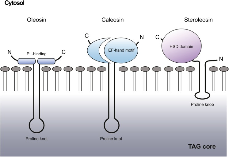
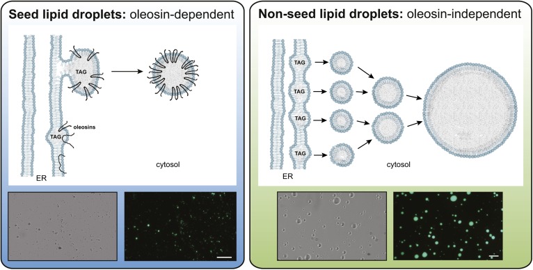
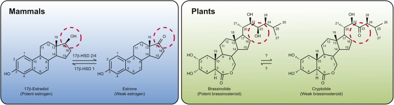
The compartmentation of neutral lipids in plants is mostly associated with seed tissues, where triacylglycerols (TAGs) stored within lipid droplets (LDs) serve as an essential physiological energy and carbon reserve during postgerminative growth. However, some nonseed tissues, such as leaves, flowers and fruits, also synthesize and store TAGs, yet relatively little is known about the formation or function of LDs in these tissues. Characterization of LD-associated proteins, such as oleosins, caleosins, and sterol dehydrogenases (steroleosins), has revealed surprising features of LD function in plants, including stress responses, hormone signaling pathways, and various aspects of plant growth and development. Although oleosin and caleosin proteins are specific to plants, LD-associated sterol dehydrogenases also are present in mammals, and in both plants and mammals these enzymes have been shown to be important in (steroid) hormone metabolism and signaling. In addition, several other proteins known to be important in LD biogenesis in yeasts and mammals are conserved in plants, suggesting that at least some aspects of LD biogenesis and/or function are evolutionarily conserved.


Figures

Fig. 1.
Fig. 1.
Schematic representations of the structures of oleosin, caleosin, and steroleosin at the surface of a TAG-filled LD. Shown are the cytosolic-facing, N- and C-terminal domains for oleosin [including the two regions of the protein proposed to be involved in its interaction with the charged, phospholipid (PL) head groups] (123), caleosin (including its calcium-binding EF-hand motif) and steroleosin (including its HSD domain). Shown also for each protein is its major hydrophobic domain, each of which is depicted as penetrating into the TAG-filled core of the LD and includes a so-called “proline knot” or “proline knob”. Based on illustrations presented in (35, 47, 74).
Fig. 2.
Fig. 2.
Schematic representation of models for oleosin-dependent and oleosin-independent LD formation from the ER in plant cells. In seed tissues (left panel), oleosins are cotranslationally inserted into the ER where they partition into domains in which TAG is accumulating between two leaflets. This promotes orientation of the oleosin proteins with N- and C termini facing the cytosol and the rest of the hydrophobic region of the protein adopting an extended hairpin configuration in the TAG matrix. The LDs in the cytosol are stabilized by oleosins and kept from fusing despite rapid dehydration and rehydration of these tissues during seed desiccation and imbibition. Micrographs are of isolated LDs from Arabidopsis seeds in bright-field (left) or by epifluorescence (right) following staining with Bodipy 493/503, a neutral lipid selective stain. LDs in nonseed tissues (right panel) may form from smaller TAG droplets that initially pinch off from the ER, then fuse to form larger droplets. Oleosins are not present in these LDs and the protein composition of LDs in nonseed tissues remains unknown. Micrographs are of LDs isolated from the oleaginous mesocarp of avocado fruit imaged in bright-field (left) or by Bodipy493/503 fluorescence (right). The white bars represent 50 microns. LDs from seeds tend to be smaller and more uniform compared with those from nonseed tissues, which fuse readily even in solution. Figure prepared by Dr. Charlene Case and Mr. Patrick Horn.
Fig. 3.
Fig. 3.
Structure and interconversion of steroid hormones in animals and plants. In mammals, 17β-estradiol, a potent estrogen-type hormone that contains a hydroxyl group at the 17th position (circled), is dehydrogenated by 17 β-HSD 2/4 to produce a less active ketone-containing estrone, whereas 17 β-HSD 1 can catalyze the reverse reaction. In an analogous manner, in plants, the 23-hydroxyl group of BL (circled) can be dehydrogenated to produce a ketone-containing derivative called cryptolide. The enzyme(s) responsible for this interconversion, however, is not known.

References

    1. Chapman K. D., Ohlrogge J. 2011. TAG accumulation in plants. J. Biol. Chem. In press. - PMC - PubMed
    1. Ghosh A. K., Chauhan N., Rajakumari S., Daum G., Rajasekharan R. 2009. At4g24160, a soluble acyl-coenzyme A-dependent lysophosphatidic acid acyltransferase. Plant Physiol. 151: 869–881. - PMC - PubMed
    1. James C. N., Horn P. J., Case C. R., Gidda S. K., Zhang D., Mullen R. T., Dyer J. M., Anderson R. G., Chapman K. D. 2010. Disruption of the Arabidopsis CGI-58 homologue produces Chanarin-Dorfman-like lipid droplet accumulation in plants. Proc. Natl. Acad. Sci. USA. 107: 17833–17838. - PMC - PubMed
    1. Murphy D. J. The dynamic roles of intracellular lipid droplets: from archaea to mammals. Protoplasma. Epub ahead of print. October 15, 2011. - PubMed
    1. Frey-Wyssling A., Grieshaber E., Mühlethaler K. 1963. Origin of spherosomes in plant cells. J. Ultrastruct. Res. 8: 506–516.

Publication types