Clinicopathological significance of altered Notch signaling in extrahepatic cholangiocarcinoma and gallbladder carcinoma
- PMID: 22046092
- PMCID: PMC3199562
- DOI: 10.3748/wjg.v17.i35.4023
Clinicopathological significance of altered Notch signaling in extrahepatic cholangiocarcinoma and gallbladder carcinoma
Abstract
Aim: To investigate the role and clinicopathological significance of aberrant expression of Notch receptors and Delta-like ligand-4 (DLL4) in extrahepatic cholangiocarcinoma and gallbladder carcinoma.
Methods: One hundred and ten patients had surgically resected extrahepatic cholangiocarcinoma (CC) and gallbladder carcinoma specimens examined by immunohistochemistry of available paraffin blocks. Immunohistochemistry was performed using anti-Notch receptors 1-4 and anti-DLL4 antibodies. We scored the immunopositivity of Notch receptors and DLL4 expression by percentage of positive tumor cells with cytoplasmic expression and intensity of immunostaining. Coexistent nuclear localization was evaluated. Clinicopathological parameters and survival data were compared with the expression of Notch receptors 1-4 and DLL4.
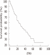
Results: Notch receptor proteins showed in the cytoplasm with or without nuclear expression in cancer cells, as well as showing weak cytoplasmic expression in non-neoplastic cells. By semiquantitative evaluation, positive immunostaining of Notch receptor 1 was detected in 96 cases (87.3%), Notch receptor 2 in 97 (88.2%), Notch receptor 3 in 97 (88.2%), Notch receptor 4 in 103 (93.6), and DLL4 in 84 (76.4%). In addition, coexistent nuclear localization was noted [Notch receptor 1; 18 cases (18.8%), Notch receptor 2; 40 (41.2%), Notch receptor 3; 32 (33.0%), Notch receptor 4; 99 (96.1%), DLL4; 48 (57.1%)]. Notch receptor 1 expression was correlated with advanced tumor, node, metastasis (TNM) stage (P = 0.043), Notch receptor 3 with advanced T stage (P = 0.017), tendency to express in cases with nodal metastasis (P = 0.065) and advanced TNM stage (P = 0.052). DLL4 expression tended to be related to less histological differentiation (P = 0.095). Coexistent nuclear localization of Notch receptor 3 was related to no nodal metastasis (P = 0.027) and Notch receptor 4 with less histological differentiation (P = 0.036), while DLL4 tended to be related inversely with T stage (P = 0.053). Coexistent nuclear localization of DLL4 was related to poor survival (P = 0.002).
Conclusion: Aberrant expression of Notch receptors 1 and 3 play a role during cancer progression, and cytoplasmic nuclear coexistence of DLL4 expression correlates with poor survival in extrahepatic CC and gallbladder carcinoma.
Keywords: Cholangiocarcinoma; Delta-like ligand-4; Gallbladder carcinoma; Immunohistochemistry; Notch receptors.
Figures


References
-
- de Groen PC, Gores GJ, LaRusso NF, Gunderson LL, Nagorney DM. Biliary tract cancers. N Engl J Med. 1999;341:1368–1378. - PubMed
-
- Welzel TM, McGlynn KA, Hsing AW, O’Brien TR, Pfeiffer RM. Impact of classification of hilar cholangiocarcinomas (Klatskin tumors) on the incidence of intra- and extrahepatic cholangiocarcinoma in the United States. J Natl Cancer Inst. 2006;98:873–875. - PubMed
-
- Berthiaume EP, Wands J. The molecular pathogenesis of cholangiocarcinoma. Semin Liver Dis. 2004;24:127–137. - PubMed
-
- Shi W, Harris AL. Notch signaling in breast cancer and tumor angiogenesis: cross-talk and therapeutic potentials. J Mammary Gland Biol Neoplasia. 2006;11:41–52. - PubMed
MeSH terms
Substances
LinkOut - more resources
Full Text Sources
Other Literature Sources
Medical

