. 2011 Oct;7(10):e1002339.
doi: 10.1371/journal.ppat.1002339. Epub 2011 Oct 27.

Coordinated destruction of cellular messages in translation complexes by the gammaherpesvirus host shutoff factor and the mammalian exonuclease Xrn1

Affiliations

Coordinated destruction of cellular messages in translation complexes by the gammaherpesvirus host shutoff factor and the mammalian exonuclease Xrn1

Sergio Covarrubias et al. PLoS Pathog. 2011 Oct.

Abstract

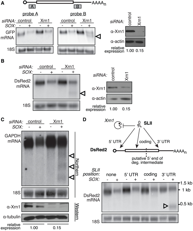
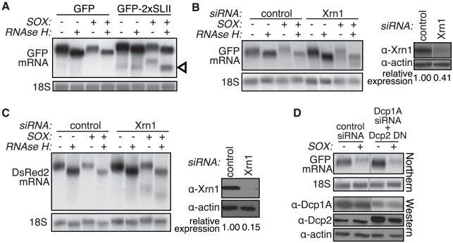
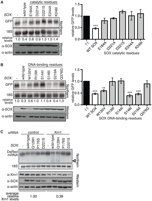
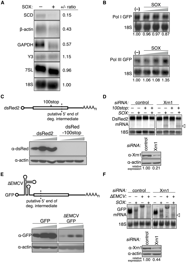
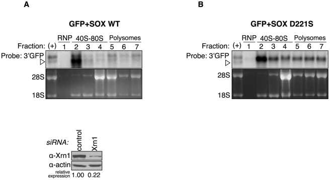
Several viruses encode factors that promote host mRNA degradation to silence gene expression. It is unclear, however, whether cellular mRNA turnover pathways are engaged to assist in this process. In Kaposi's sarcoma-associated herpesvirus this phenotype is enacted by the host shutoff factor SOX. Here we show that SOX-induced mRNA turnover is a two-step process, in which mRNAs are first cleaved internally by SOX itself then degraded by the cellular exonuclease Xrn1. SOX therefore bypasses the regulatory steps of deadenylation and decapping normally required for Xrn1 activation. SOX is likely recruited to translating mRNAs, as it cosediments with translation initiation complexes and depletes polysomes. Cleaved mRNA intermediates accumulate in the 40S fraction, indicating that recognition occurs at an early stage of translation. This is the first example of a viral protein commandeering cellular mRNA turnover pathways to destroy host mRNAs, and suggests that Xrn1 is poised to deplete messages undergoing translation in mammalian cells.


Conflict of interest statement

The authors have declared that no competing interests exist.

Figures

Figure 1
Figure 1. Xrn1 is required for the removal of a 3′ intermediate during SOX-mediated mRNA degradation.
(A–B) 293T cells were treated with control or Xrn1 siRNAs, then transfected with the indicated reporters +/− SOX. The level of Xrn1 protein depletion is shown on the right, with actin as a loading control. (A) Diagram depicts location of probes on the GFP message. Northern blots using probes against either the 5′ (probe A; left panel) or 3′ (probe B; right panel) UTR of the GFP mRNA reporter or 18S. (B) Northern blots using probes against the 3′ UTR of the DsRed2 mRNA reporter, or 18S (left panels). (C) 293T cells were treated with control or Xrn1 shRNAs, then transfected with GFP (−) or GFP-SOX (+). Cells were then selected for GFP fluorescence by FACS before RNA and protein collection. RNA was Northern blotted with probes to the 3′ end of the GAPDH coding region, or to 18S. The level of Xrn1 knockdown was assessed by Western blot, using tubulin as a loading control. In panels A, B and C, arrowheads denote degradation intermediates. (D) A flaviviral Xrn1-blocking element (SLII) was inserted at different positions within the DsRed2 mRNA. RNA from 293T cells transfected with the indicated reporter +/− SOX was Northern blotted using a probe directed against the 3′ UTR of the DsRed2 reporter, or 18S. The full-length mRNA is ∼1.2 kb, and the expected size of the fragment protected from Xrn1 degradation by SLII within the 5′ UTR is ∼1.1 kb, within the coding region is ∼900 bp, and within the 3′ UTR is ∼500 bp. Arrowhead denotes protected fragment.
Figure 2
Figure 2. The 3′ degradation intermediate retains its poly(A) tail.
(A) 293T cells were transfected with GFP or a modified GFP reporter containing two copies of the flaviviral Xrn1-blocking element SLII within the GFP coding region, +/− SOX. A fraction of the RNA was treated with oligo(dT) and RNAse H to remove the poly(A) tail prior to Northern blotting with a GFP 3′ UTR or an 18S probe. Arrowhead denotes protected fragments. (B–C) 293T cells were transfected with control or Xrn1 siRNAs, followed by GFP (B) or DsRed2 (C) reporters +/− SOX. A fraction of the RNA was treated with oligo(dT) and RNAse H to remove the poly(A) tail prior to Northern blotting with a GFP 3′ UTR or an 18S probe. Xrn1 protein levels were assessed by Western blot (right panels in B and C). (D) 293T cells were treated with control siRNAs or siRNAs against the decapping complex protein Dcp1A. They were then transfected with GFP +/− SOX and/or the Dcp2 dominant negative mutant E148Q (Dcp2 DN). RNA was Northern blotted using a GFP 3′ UTR or an 18S probe. Western blots show the level of Dcp1A knockdown and Dcp2 DN overexpression. Actin serves as a loading control and grey lines indicate where intervening lanes have been cropped out. See also Figure S2.
Figure 3
Figure 3. A specific element in the GFP coding region is sufficient for production of degradation intermediates by SOX.
(A) The modified GFP mRNA (GFPrep) contains an internal repeat of the first 201 basepairs of the GFP coding region (grey shaded area). (B) 293T cells were treated with control or Xrn1 siRNAs, then transfected with GFP or GFPrep +/− SOX. RNA was Northern blotted with a GFP 3′ UTR or an 18S probe. Arrowheads denote degradation intermediates. (C) Western blot shows the level of Xrn1 depletion in the experiment depicted in panel B, with actin as a loading control. (D) 5′ RACE was carried out on total RNA from 293T cells transfected with Xrn1 siRNAs and a GFP expression vector in the presence or absence of SOX. An internal GFP primer and a primer to the adapter ligated to the 5′ end of the RNAs were used to amplify GFP in each sample. Full length refers to the band corresponding to the intact GFP transcript, intermediate refers to the ∼300 bp band preferentially amplified in the lysate from SOX-expressing cells, and NT denotes the no template control. (E) A conserved TGAAG site located just upstream of the cleavage site was deleted from GFP to create GFP-ΔTGAAG. 293T cells were treated with control or Xrn1 siRNAs, then transfected with GFP or GFP ΔTGAAG in the presence or absence of SOX. RNA was Northern blotted with a GFP 3′ UTR or 18S probe (upper panels). Arrowheads denote degradation intermediates. Western blot (lower panels) shows the level of Xrn1 depletion in experiments depicted in panel E and F, with tubulin as a loading control. (F) Modified GFP reporters bearing an internal duplication of sequences around the GFP cleavage site were transfected in 293T cells treated with Xrn1 siRNAs +/− SOX. The inserted nucleotides are numbered based on their position relative to the GFP start codon. GFPrep (nt 1–201) is included as a positive control. RNA was Northern blotted with a GFP 3′ UTR or 18S probe. Arrowheads denote degradation intermediates.
Figure 4
Figure 4. The catalytic residues of SOX are required for mRNA turnover and generation of the degradation intermediate.
(A–B) Wild-type (WT) SOX or SOX putative catalytic (A) or DNA binding (B) residue mutants were transfected into 293T cells with a GFP reporter. RNA was Northern blotted using a 5′ GFP or 18S probe, and protein lysates were Western blotted for SOX and actin (as a loading control). Graphs depicting mean relative GFP levels ± s.e.m. from >3 experiments in cells transfected with the various mutants are also shown (right). **p<0.01, ***p<0.001, One-way ANOVA followed by Dunnett's test versus (−). (C) 293T cells were transfected with control or Xrn1 siRNAs, followed by a DsRed2 reporter +/− WT SOX or the indicated SOX mutant. Northern blots were performed using a probe directed against the 3′ UTR of the DsRed2 mRNA or 18S (upper panels), and Western blots show expression of the SOX mutants and the level of Xrn1 depletion (lower panels). Arrowhead denotes degradation intermediates.
Figure 5
Figure 5. SOX depletes polyribosomes and cosediments with 40S subunits.
(A) TREx BCBL-1-RTA (BCBL-1) cells were mock treated (latent) or induced (lytic) with 1 µg/ml doxycycline, 500 ng/ml ionomycin, and 20 ng/ml 2-O-tetradecanoylphorbol-13-acetate for 24 h to stimulate KSHV replication, then subjected to sucrose gradient fractionation using a 15–60% sucrose gradient in order to monitor the abundance of translating polysomes. (B) Fractions collected from the induced BCBL-1 gradients shown in panel A were Western blotted with the indicated antibodies. (C) Lysate from induced BCBL-1 cells was fractionated using a 5–20% sucrose gradient and analyzed by Western blot with indicated antibodies. (D) 293T cells were transfected with the indicated construct and subjected to sucrose gradient fractionation to obtain polysome profiles. Dashed lines indicate polysome levels of either latent cells (A) or vector expressing cells (D).
Figure 6
Figure 6. SOX targets mRNA at an early stage of translation.
(A) 293T cells were transfected with control or Xrn1 shRNA-expressing constructs, and subsequently with either GFP or GFP-SOX. Cells were then sorted for GFP fluorescence to generate a pure population of SOX-expressing and control cells prior to RNA extraction. RNA was Northern blotted with the indicated probes against endogenous Pol I (18S), Pol II (SCD, β-actin, GAPDH), and Pol III (Y3, 7SL) RNAs. (B) 293T cells were transfected with either Pol I-driven GFP or Pol III-driven GFP reporters with or without increasing amounts of SOX (200–600 ng), then total RNA was Northern blotted using GFP or 18S probes. (C) 293T cells were transfected with increasing amounts (100–300 ng) of dsRed2 or a dsRed2–100stop construct containing a premature termination codon upstream of the predicted cleavage site (dashed line), then Western blotted for dsRed protein. (D) 293T cells were transfected with control or Xrn1 shRNAs and subsequently with either the dsRed2 or dsRed2-100stop (100stop) reporter in the presence or absence of SOX. RNA was Northern blotted with 3′ end dsRed2 or 18S probes. The arrowhead indicates the position of the SOX-induced cleavage product. (E) 293T cells were transfected with increasing amounts (100–300 ng) of GFP or a ΔEMCV-GFP construct containing the ΔEMCV IRES in the 5′ UTR, then Western blotted for GFP protein. (F) 293T cells were transfected with control or Xrn1 shRNAs and then with either the GFP or ΔEMCV-GFP reporter in the presence or absence of SOX. RNA was Northern blotted with 3′ GFP or 18S probes. The arrowhead indicates the position of the SOX-induced cleavage product. The Western blot shows level of Xrn1 depletion, and the actin loading control.
Figure 7
Figure 7. The degradation intermediate sediments predominantly with the 40S fractions.
293T cells were transfected with Xrn1 shRNA, followed by expression of GFP and either wild-type (WT) SOX (A) or the catalytically dead SOX D221S (B). They were then fractionated over sucrose gradients and RNA from each fraction was Northern blotted with a 3′ GFP probe. Ribosomal RNA was visualized by ethidium bromide staining. In both panels the (+) lane shows the migration of full-length GFP and the degradation intermediate in unfractionated RNA from cells expressing wild-type SOX. It was used as a reference to identify the different RNA species in the fractionated RNA. Arrowheads point to degradation intermediates.

References

    1. Graham JR, Hendershott MC, Terragni J, Cooper GM. mRNA degradation plays a significant role in the program of gene expression regulated by phosphatidylinositol 3-kinase signaling. Mol Cell Biol. 2010;30:5295–5305. - PMC - PubMed
    1. Fan J, Yang X, Wang W, Wood WH, 3rd, Becker KG, et al. Global analysis of stress-regulated mRNA turnover by using cDNA arrays. Proc Natl Acad Sci U S A. 2002;99:10611–10616. - PMC - PubMed
    1. Cheadle C, Fan J, Cho-Chung YS, Werner T, Ray J, et al. Control of gene expression during T cell activation: alternate regulation of mRNA transcription and mRNA stability. BMC Genomics. 2005;6:75. - PMC - PubMed
    1. Belasco JG. All things must pass: contrasts and commonalities in eukaryotic and bacterial mRNA decay. Nat Rev Mol Cell Biol. 2010;11:467–478. - PMC - PubMed
    1. Garneau NL, Wilusz J, Wilusz CJ. The highways and byways of mRNA decay. Nat Rev Mol Cell Biol. 2007;8:113–126. - PubMed

Publication types

MeSH terms