

Immune Responses of HIV-1 Tat Transgenic Mice to Mycobacterium Tuberculosis W-Beijing SA161
- PMID: 22046211
- PMCID: PMC3204420
- DOI: 10.2174/1874613601105010086
Immune Responses of HIV-1 Tat Transgenic Mice to Mycobacterium Tuberculosis W-Beijing SA161
Abstract
Background: Mycobacterium tuberculosis remains among the leading causes of death from an infectious agent in the world and exacerbates disease caused by the human immunodeficiency virus (HIV). HIV infected individuals are prone to lung infections by a variety of microbial pathogens, including M. tuberculosis. While the destruction of the adaptive immune response by HIV is well understood, the actual pathogenesis of tuberculosis in co-infected individuals remains unclear. Tat is an HIV protein essential for efficient viral gene transcription, is secreted from infected cells, and is known to influence a variety of host inflammatory responses. We hypothesize Tat contributes to pathophysiological changes in the lung microenvironment, resulting in impaired host immune responses to infection by M. tuberculosis.
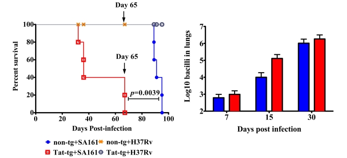
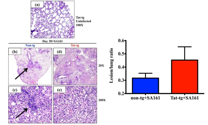
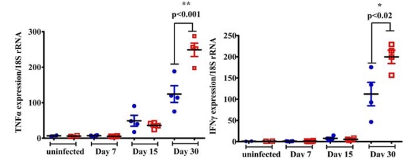
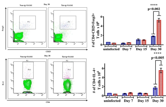
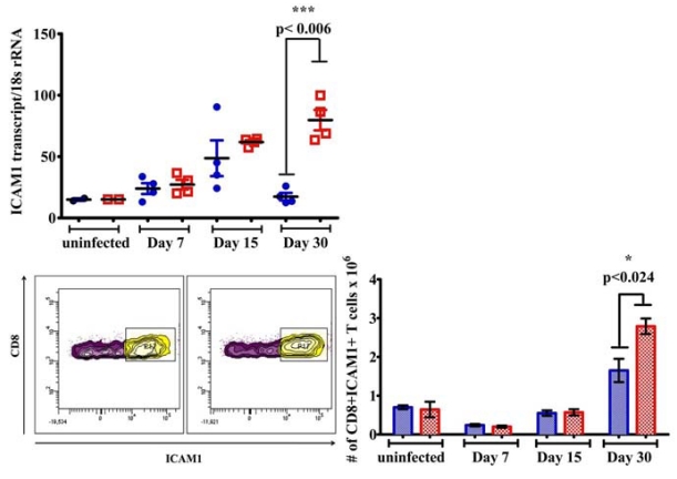
Results: Herein, we show transgenic mice that express Tat by lung alveolar cells are more susceptible than non-transgenic control littermates to a low-dose aerosol infection of M. tuberculosis W-Beijing SA161. Survival assays demonstrate accelerated mortality rates of the Tat transgenic mice compared to non-transgenics. Tat transgenic mice also showed poorly organized lung granulomata-like lesions. Analysis of the host immune response using quantitative RT-PCR, flow cytometry for surface markers, and intracellular cytokine staining showed increased expression of pro-inflammatory cytokines in the lungs, increased numbers of cells expressing ICAM1, increased numbers of CD4+CD25+Foxp3+ T regulatory cells, and IL-4 producing CD4+ T cells in the Tat transgenics compared to infected non-tg mice.
Conclusions: Our data show quantitative differences in the inflammatory response to the SA161 clinical isolate of M. tuberculosis W-Beijing between Tat transgenic and non-transgenic mice, suggesting Tat contributes to the pathogenesis of tuberculosis.
Keywords: HIV-1; Mycobacterium tuberculosis W-Beijing; Tat; immune responses..
Figures
Similar articles
-
HIV-1 Tat protein vaccination in mice infected with Mycobacterium tuberculosis is safe, immunogenic and reduces bacterial lung pathology.BMC Infect Dis. 2016 Aug 22;16(1):442. doi: 10.1186/s12879-016-1724-7. BMC Infect Dis. 2016. PMID: 27549342 Free PMC article.
-
Human IL-32 expression protects mice against a hypervirulent strain of Mycobacterium tuberculosis.Proc Natl Acad Sci U S A. 2015 Apr 21;112(16):5111-6. doi: 10.1073/pnas.1424302112. Epub 2015 Mar 27. Proc Natl Acad Sci U S A. 2015. PMID: 25820174 Free PMC article.
-
Altered cytokine expression in T lymphocytes from human immunodeficiency virus Tat transgenic mice.J Virol. 1995 Dec;69(12):7622-9. doi: 10.1128/JVI.69.12.7622-7629.1995. J Virol. 1995. PMID: 7494270 Free PMC article.
-
[Novel vaccines against M. tuberculosis].Kekkaku. 2006 Dec;81(12):745-51. Kekkaku. 2006. PMID: 17240920 Review. Japanese.
-
Evolutionary Genetics of Mycobacterium tuberculosis and HIV-1: "The Tortoise and the Hare".Microorganisms. 2021 Jan 11;9(1):147. doi: 10.3390/microorganisms9010147. Microorganisms. 2021. PMID: 33440808 Free PMC article. Review.
Cited by
-
HIV-1 Tat protein vaccination in mice infected with Mycobacterium tuberculosis is safe, immunogenic and reduces bacterial lung pathology.BMC Infect Dis. 2016 Aug 22;16(1):442. doi: 10.1186/s12879-016-1724-7. BMC Infect Dis. 2016. PMID: 27549342 Free PMC article.
-
The chosen few: Mycobacterium tuberculosis isolates for IMPAc-TB.Front Immunol. 2024 Oct 28;15:1427510. doi: 10.3389/fimmu.2024.1427510. eCollection 2024. Front Immunol. 2024. PMID: 39530100 Free PMC article. Review.
-
The Impact of Animal Models and Strain Standardization on the Evaluation of Tuberculosis Vaccine Efficacy.Vaccines (Basel). 2025 Jun 21;13(7):669. doi: 10.3390/vaccines13070669. Vaccines (Basel). 2025. PMID: 40733646 Free PMC article. Review.
References
-
- Granich R, Akolo C, Gunneberg C, et al. Prevention of tuberculosis in people living with HIV. Clin Infect Dis. 2010;50(Suppl 3):S215–22. - PubMed
-
- Kitaura H, Ohara N, Kobayashi K, et al. TNF-alpha-mediated multiplication of human immunodeficiency virus in chronically infected monocytoid cells by mycobacterial infection. APMIS. 2001;109(7-8):533–40. - PubMed
-
- Lederman MM, Georges DL, Kusner DJ, et al. Mycobacterium tuberculosis and its purified protein derivative activate expression of the human immunodeficiency virus. J Acquir Immune Defic Syndr. 1994;7(7):727–33. - PubMed
Grants and funding
LinkOut - more resources
Full Text Sources
Research Materials
Miscellaneous