. 2011;6(10):e26543.
doi: 10.1371/journal.pone.0026543. Epub 2011 Oct 27.

Expression of sympathetic nervous system genes in Lamprey suggests their recruitment for specification of a new vertebrate feature

Affiliations

Expression of sympathetic nervous system genes in Lamprey suggests their recruitment for specification of a new vertebrate feature

Daniela Häming et al. PLoS One. 2011.

Abstract

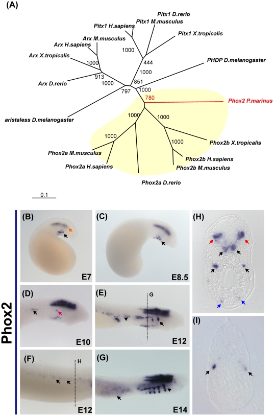
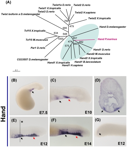
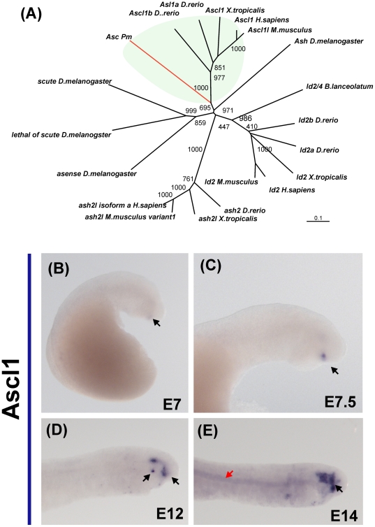
The sea lamprey is a basal, jawless vertebrate that possesses many neural crest derivatives, but lacks jaws and sympathetic ganglia. This raises the possibility that the factors involved in sympathetic neuron differentiation were either a gnathostome innovation or already present in lamprey, but serving different purposes. To distinguish between these possibilities, we isolated lamprey homologues of transcription factors associated with peripheral ganglion formation and examined their deployment in lamprey embryos. We further performed DiI labeling of the neural tube combined with neuronal markers to test if neural crest-derived cells migrate to and differentiate in sites colonized by sympathetic ganglia in jawed vertebrates. Consistent with previous anatomical data in adults, our results in lamprey embryos reveal that neural crest cells fail to migrate ventrally to form sympathetic ganglia, though they do form dorsal root ganglia adjacent to the neural tube. Interestingly, however, paralogs of the battery of transcription factors that mediate sympathetic neuron differentiation (dHand, Ascl1 and Phox2b) are present in the lamprey genome and expressed in various sites in the embryo, but fail to overlap in any ganglionic structures. This raises the intriguing possibility that they may have been recruited during gnathostome evolution to a new function in a neural crest derivative.


Conflict of interest statement

Competing Interests: The authors have declared that no competing interests exist.

Figures

Figure 1
Figure 1. DiI labeling of lamprey neural crest cells reveals absence of sympathetic ganglia during embryonic development.
(A) Focal injections of DiI in the lamprey neural tube at day 5 results in labeling of migrating cephalic neural crest (arrow in B). However, focal injections into the posterior neural tube (C) fail to label trunk neural crest cells. D) Filling the lumen of the neural tube with DiI after cavitation produces labeled trunk neural crest cells in several neural crest derivatives (E). A section through an injected embryo (F) shows labeling of the dorsal root ganglia (DRG, arrow in G), the mesenchyme of the fin (H) and neurons surrounding the gut (F), but no structure that resembles sympathetic ganglia. Neurofilament staining (J) labels neural crest derivatives such as the DRG but also fails to reveal any sympathetic like structures.
Figure 2
Figure 2. Expression of the transcription factor Phox2 in embryos of P. marinus.
(A) Phylogenetic analysis of a putative Phox2 fragment places it at the base of the gnathostome Phox2a and Phox2b gene families. (B) Expression of Phox2 is initially observed on the hindbrain (red arrow) and on a group of cells of the ventral mesenchyme (black arrow). (C) This expression pattern is maintained at day 8.5, as Phox2 positive cells migrate ventrally (black arrow). (D) At day 10, a stream of positive cells is observed above the heart and appears to be migrating posteriorly (black arrow). At this stage, Phox2 transcripts are first detected on the epibranchial ganglia (red arrow). (E) At day 21, the expression domain of Phox2 expands to include cranial (black arrow) and epibranchial ganglia (arrowhead). A section of this embryo (H) reveals staining in the motor neurons of the hindbrain, epibranchial ganglia and ventral mesenchyme (red, black and blue arrows, respectively). (F) At posterior axial levels, Phox2b expressing cells are observed adjacent to the yolk sac (section on I). (G) At later stages, a larger number of cells around the yolk start expressing Phox2 (black arrow), and there is strong expression in cranial nerves (arrowhead).
Figure 3
Figure 3. Expression of the helix-loop-helix transcription factor Hand in lamprey embryos.
(A) Phylogenetic analysis suggests lamprey has one ortholog of both the dHand and eHand gnathostome genes. (B) Lamprey Hand expression is first observed in the cardiac field, and is conspicuous after 7 days of development. (B) At day 10, two domains of expression are clearly present: the heart (black arrow) and a part of the anterior mesenchyme (red arrow). (C) Expression in the cardiac ganglia (black arrow) is first detected at day 12 days; transverse sections reveal high abundance of transcript in the mesenchyme flanking the pharynx (black arrows on F). (E) Strong expression is observed in the ventral mesenchyme of the lamprey head (read arrow), as well as the heart at day 14 (black arrow). (G) Posterior expression appears to be restricted to the unsegmented mesoderm of the tail (black arrow).
Figure 4
Figure 4. Expression of Ascl1 during embryonic development of the lamprey.
(A) Phylogenetic analysis places the putative lamprey Ascl1 ortholog with gnathostome Ascl1 genes. (B) Onset of Ascl1 expression is apparent at day 7, when transcripts are detected in the anterior lip mesoderm. Shortly afterwards (C), faint expression is observed in the lens (arrow). (D) On day 12, lens expression is elevated (arrow), and staining on the hypophysis and trigeminal ganglia (red arrow) are also observed. (E) Finally, on day 14, Ascl1 expression is observed in the notochord (red arrow).

Similar articles

Cited by

References

    1. Gess RW, Coates MI, Rubidge BS. A lamprey from the Devonian period of South Africa. Nature. 2006;443:981–984. - PubMed
    1. Janvier P. Palaeontology: modern look for ancient lamprey. Nature. 2006;443:921–924. - PubMed
    1. Kuraku S, Hoshiyama D, Katoh K, Suga H, Miyata T. Monophyly of lampreys and hagfishes supported by nuclear DNA-coded genes. Journal of molecular evolution. 1999;49:729–735. - PubMed
    1. Osorio J, Retaux S. The lamprey in evolutionary studies. Development Genes and Evolution. 2008;218:221–235. - PubMed
    1. Le Douarin N. Cambridge University Press; 1982. The neural crest.

Publication types

Substances