. 2011;6(10):e26789.
doi: 10.1371/journal.pone.0026789. Epub 2011 Oct 28.

N-cofilin can compensate for the loss of ADF in excitatory synapses

Affiliations

N-cofilin can compensate for the loss of ADF in excitatory synapses

Andreas Görlich et al. PLoS One. 2011.

Abstract

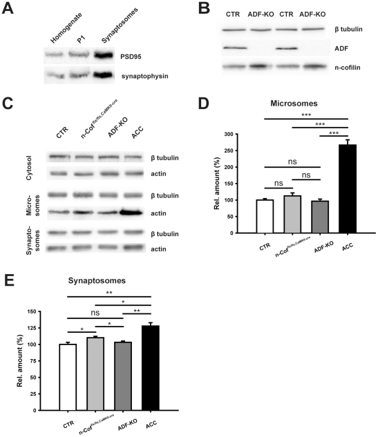
Actin plays important roles in a number of synaptic processes, including synaptic vesicle organization and exocytosis, mobility of postsynaptic receptors, and synaptic plasticity. However, little is known about the mechanisms that control actin at synapses. Actin dynamics crucially depend on LIM kinase 1 (LIMK1) that controls the activity of the actin depolymerizing proteins of the ADF/cofilin family. While analyses of mouse mutants revealed the importance of LIMK1 for both pre- and postsynaptic mechanisms, the ADF/cofilin family member n-cofilin appears to be relevant merely for postsynaptic plasticity, and not for presynaptic physiology. By means of immunogold electron microscopy and immunocytochemistry, we here demonstrate the presence of ADF (actin depolymerizing factor), a close homolog of n-cofilin, in excitatory synapses, where it is particularly enriched in presynaptic terminals. Surprisingly, genetic ablation of ADF in mice had no adverse effects on synapse structure or density as assessed by electron microscopy and by the morphological analysis of Golgi-stained hippocampal pyramidal cells. Moreover, a series of electrophysiological recordings in acute hippocampal slices revealed that presynaptic recruitment and exocytosis of synaptic vesicles as well as postsynaptic plasticity were unchanged in ADF mutant mice. The lack of synaptic defects may be explained by the elevated n-cofilin levels observed in synaptic structures of ADF mutants. Indeed, synaptic actin regulation was impaired in compound mutants lacking both ADF and n-cofilin, but not in ADF single mutants. From our results we conclude that n-cofilin can compensate for the loss of ADF in excitatory synapses. Further, our data suggest that ADF and n-cofilin cooperate in controlling synaptic actin content.


Conflict of interest statement

Competing Interests: The authors have declared that no competing interests exist.

Figures

Figure 1
Figure 1. ADF is present in pre- and postsynaptic structures of excitatory synapses.
(A) Immunoblot analyses demonstrated the specificity of the ADF antibody used for expression and localization studies: no band was detectable in cortical (CX), hippocampal (HIP) or striatal (STR) protein lysates from homozygous ADF mutants (−/−). N-cofilin levels were unchanged in lysates from homozygous ADF mutants. (B) Immunoblot analysis demonstrated the broad expression of ADF in the brain throughout postnatal development (postnatal day 0 (P0) to P80). CB: cerebellum, BS: brainstem. (C) Immunocytochemistry of cultured hippocampal pyramidal cells after 21 days in culture demonstrating the presence of ADF (green) in the cell body and in neurites. F-actin-rich structures were visualized with fluorescent-labeled phalloidin (red). Scale bar: 50 µm. (D) High magnification of a dendritc shaft, demonstrating the presence of ADF in phalloidin-positive, F-actin-rich structures such as dendritic spines. Scale bar: 5 µm. (E) Co-labeling with synaptophysin (synapto; red) revealed the presence of ADF (green) in presynaptic structures. Scale bar: 5 µm. (F) Likewise, ADF (green) co-localized with PSD-95 (red), a marker of the postsynaptic density. Scale bar: 5 µm. Small images in D–F: binary masks (green or red channel) and merged masks of dendritic structure indicated by dashed boxes. (G+H) Representative electron micrographs of the CA1 and CA3 stratum radiatum demonstrate localization of ADF in presynaptic boutons (b) and dendritic spines (sp). Arrows point to exemplarily gold particles that label ADF. Scale bars: 150 nm in G, 200 nm in H. (I) In the CA1 and CA3 region, density of gold particles was significantly higher in presynaptic boutons than in dendritic spines.
Figure 2
Figure 2. Unaltered dendritic spine density and morphology in CA1 pyramidal cells.
(A) Representative examples of Golgi-stained CA1 pyramidal cells indicate similar branching and complexity of the dendritic trees in controls (CTR) and ADF mutants (ADF-KO). Scale bar: 50 µm. (B) Sholl analysis of the apical dendritic tree in the stratum radiatum revealed no difference between both genotypes (n = 8 for CTR; n = 6 for ADF-KO). (C) Representative images of 2nd order dendritic branches from Golgi-stained pyramidal cells in the stratum radiatum of CA1. Scale bar: 2.5 µm. (D) The total number of spines (CTR: 27.1±0.2 spines/20 µm, ADF-KO: 27.9±0.5 spines/20 µm, P = 0.715) as well as the numbers of mushroom-like (CTR: 14.2±0.2 spines/20 µm, ADF-KO: 15.0±0.2 spines/20 µm, P = 0.540), stubby (CTR: 7.1±0.1 spines/20 µm, ADF-KO: 6.5±0.1 spines/20 µm, P = 0.435) and thin spines were unaltered in ADF-KO (CTR: 5.8±0.1 spines/20 µm, ADF-KO: 6.3±0.1 spines/20 µm, P = 0.583; more than 1,000 µm dendritic length was analyzed in four mice per genotype). (E+F) Representative electron micrographs of CA1 stratum radiatum from a CTR and an ADF-KO mouse. Image size: 2.27 µm2; b: presynaptic bouton, sp: postsynaptic spine, *: postsynaptic density. (G) Spine area (CTR: 0.098±0.004 µm2, n = 181 spines from 3 mice; ADF-KO: 0.102±0.004 µm2, n = 185/3; P = 0.512) and (H) PSD length (CTR: 0.193±0.005 µm, n = 184 spines from 3 mice; ADF-KO: 0.201±0.005 µm, n = 183/3; P = 0.277) were unchanged in ADF-KO as deduced from the cumulative distribution curves and the mean values (insets).
Figure 3
Figure 3. Basal synaptic transmission and presynaptic physiology were independent of ADF.
(A) Input-output curves were not different between CTR and ADF-KO (n = 12 for CTR; n = 16 for ADF-KO), indicating unchanged basal synaptic transmission in ADF-KO. (B) Paired pulse ratio (PPR) at different interstimulus intervals (10 to 200 ms) was unaltered in ADF-KO (n = 12 for CTR; n = 18 for ADF-KO). (C) Exemplary traces showing miniature excitatory postsynaptic currents (mEPSC) recorded from a CTR (upper trace) and an ADF-KO pyramidal cell (lower trace). (D+E) Both mEPSC amplitudes and interevent intervals (IEI) were unaltered in ADF-KO as deduced from the cumulative curves and mean values (insets; mEPSC amplitudes: CTR: 7.29±0.33 pA, n = 14 cells from 7 mice; ADF-KO: 7.09±0.42, n = 9/3; P = 0.699; IEI: CTR: 0.831±0.049 s; ADF-KO: 0.712±0.031 s; P = 0.089) were unaltered in ADF-KO. (F) Additionally, presynaptic short-term vesicle depression induced by 10 Hz stimulation for 2 min was similar in CTR and ADF-KO.
Figure 4
Figure 4. ADF is dispensable for synaptic plasticity.
(A) Long-term depression induced by low frequency stimulation (1 Hz for 15 min) with paired pulses (interpulse interval 50 ms) showed no difference between CTR and ADF-KO (last 10 min of recording; CTR: 0.725±0.042, n = 8; ADF-KO: 0.706±0.038, n = 9; P = 0.739). (B) Likewise, long-term potentiation induced by a single tetanic stimulation with 100 Hz for 1 s was indistinguishable between CTR and ADF-KO (last 10 min of recording; CTR: 1.205±0.035, n = 12; ADF-KO: 1.221±0.028, n = 12; P = 0.732).
Figure 5
Figure 5. Normal learning and memory in ADF-KO.
(A) Freezing behavior in a fear-conditioning paradigm during the context (CTR: 23.4±3.0%, n = 17; ADF-KO: 21.0±2.8, n = 15; P = 0.577) and cue testing (CTR: 32.3±5.8%; ADF-KO: 33.8±5.4; P = 0.848) was unchanged in ADF-KO. HAB: habituation, CS: conditioned stimulus. (B–D) Likewise, a two-choice serial reaction task revealed no learning defect in ADF-KO. (B) The number of operative responses as well as (C) the latency for reaching the maximum of 50 operative responses or (D) the ratio of correct versus incorrect responses were unchanged between CTR and ADF-KO (n = 8 for both genotypes).
Figure 6
Figure 6. N-cofilin compensates the loss of ADF at synapses.
(A) Immunoblot analyses show enrichment of synaptic markers (PSD-95, synaptophysin) in synaptosomes. Equal protein load was verified by Coomassie staining of SDS-PAGEs. Compared to total protein lysates (homogenate) and the organelle- and nuclei-containing fraction P1 PSD-95 and synaptophysin signals were increased in synaptosomes. (B) Immunoblots showing that synaptic n-cofilin levels were increased in synaptosomes from ADF-KO. (C) Immunoblot analysis demonstrating equal cytosolic actin levels in CTR, n-cofilin mutants (n-Cofflx/flx,CaMKII-cre), ADF-KO and double mutants (ACC) that lack both ADF and n-cofilin. Conversely, actin levels were increased in microsomal and synaptosomal preparations from ACC mice. (D) Quantification of microsomal actin levels. In microsomes from ACC mice, actin levels were significantly increased to 266.8±15.8% of CTR levels (n = 4; P<0.001) and significantly higher compared to n-Cofflx/flx,CaMKII-cre mice (P<0.001) or ADF-KO (P<0.001). No significant increase in actin levels was found in microsomes from ADF-KO or n-cofilin mutants. (E) Quantification of actin levels in synaptosomes. Actin levels in synaptosomes were significantly increased in n-cofilin mutants (+10.1±2.1%; n = 4; P<0.05) and ACC mice (+27.8±5.2%; n = 4; P<0.01). In ACC mice, synaptosomal actin levels were significantly higher than in synaptosomes from n-cofilin mutants (P<0.05) or ADF-KO (P<0.01). No increase was found in synaptosomes from ADF-KO.

References

    1. Matus A, Ackermann M, Pehling G, Byers HR, Fujiwara K. High actin concentrations in brain dendritic spines and postsynaptic densities. Proc Natl Acad Sci U S A. 1982;79:7590–7594. - PMC - PubMed
    1. Fifkova E, Delay RJ. Cytoplasmic actin in neuronal processes as a possible mediator of synaptic plasticity. J Cell Biol. 1982;95:345–350. - PMC - PubMed
    1. Landis DM, Hall AK, Weinstein LA, Reese TS. The organization of cytoplasm at the presynaptic active zone of a central nervous system synapse. Neuron. 1988;1:201–209. - PubMed
    1. Cingolani LA, Goda Y. Actin in action: the interplay between the actin cytoskeleton and synaptic efficacy. Nat Rev Neurosci. 2008;9:344–356. - PubMed
    1. Renner M, Choquet D, Triller A. Control of the postsynaptic membrane viscosity. J Neurosci. 2009;29:2926–2937. - PMC - PubMed

Publication types