. 2011;6(10):e26907.
doi: 10.1371/journal.pone.0026907. Epub 2011 Oct 27.

Rates of mutation and host transmission for an Escherichia coli clone over 3 years

Affiliations

Rates of mutation and host transmission for an Escherichia coli clone over 3 years

Peter R Reeves et al. PLoS One. 2011.

Abstract

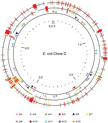
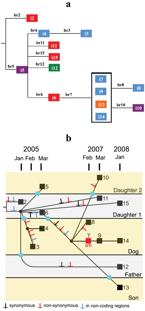
Although over 50 complete Escherichia coli/Shigella genome sequences are available, it is only for closely related strains, for example the O55:H7 and O157:H7 clones of E. coli, that we can assign differences to individual evolutionary events along specific lineages. Here we sequence the genomes of 14 isolates of a uropathogenic E. coli clone that persisted for 3 years within a household, including a dog, causing a urinary tract infection (UTI) in the dog after 2 years. The 20 mutations observed fit a single tree that allows us to estimate the mutation rate to be about 1.1 per genome per year, with minimal evidence for adaptive change, including in relation to the UTI episode. The host data also imply at least 6 host transfer events over the 3 years, with 2 lineages present over much of that period. To our knowledge, these are the first direct measurements for a clone in a well-defined host community that includes rates of mutation and host transmission. There is a concentration of non-synonymous mutations associated with 2 transfers to the dog, suggesting some selection pressure from the change of host. However, there are no changes to which we can attribute the UTI event in the dog, which suggests that this occurrence after 2 years of the clone being in the household may have been due to chance, or some unknown change in the host or environment. The ability of a UTI strain to persist for 2 years and also to transfer readily within a household has implications for epidemiology, diagnosis, and clinical intervention.


Conflict of interest statement

Competing Interests: The authors have declared that no competing interests exist.

Figures

Figure 1
Figure 1. A tree of the E. coli strains (including Shigella strains) with full genome sequences.
The phylogenetic tree was constructed based on the alignments of the core genomes of the 56 E. coli/Shigella genome sequences available (Table S9), including clone D (i2) and clone A (i1) described in this paper. The genome of E. fergusonii was used as outgroup. Bootstrap values are given at each node. The multiple genome sequences for E. coli K-12, E. coli B, and E. coli O157:H7 were each combined as a single entry.
Figure 2
Figure 2. Comparison of the genomes of clone D and CFT073, and distribution of SNPs in clone D isolates.
The outer ring represents the genome of CFT073 and the middle ring that of clone D. Both rings show insertions (red) and deletions (green) with respect to the other strain, with the distinction being determined by outgroup analysis, as described in the supporting information. Indels for which the distinction between insertion and deletion cannot be made are marked on both genomes (orange). The large indels (>10 kb) are numbered as in Table S3. The inner ring with the triangular pointers shows the SNPs found in the 14 clone D isolates. The SNPs are also numbered as in Table 2.
Figure 3
Figure 3. The relationships of the clone D isolates.
a, Phylogenetic tree of the 11 genotypes observed in clone D. Note that the tree is fully consistent with the data and outgroup analysis and requires no parallel or reverse mutations. The isolates are colour-coded for the host (Daughter 1, red; Daughter 2, dark violet; Dog, blue; Father, green; Son, orange). Branches are numbered and lengths are proportional to the number of mutations. b, Tree showing the individual isolates (squares) in relation to date of isolation. The mutations along each branch are shown (arrows). Isolates i7, i9, i13 and i14 are genetically identical, and so there are no mutations to record on their branches, which relate only to passage of time. It can be seen that two mutations (No. 1 and 2) were present in isolate i2 (January 2005), and another mutation (No. 3) was present in all other isolates, and must have arisen before February 2005, when first seen in i3. The distribution of isolation dates and hosts shows that there must have been several instances of transfer of this lineage between hosts, with the minimum number consistent with the tree data being 6, as marked on the tree (aqua circles). Black circles indicate branch points (no isolate).

References

    1. Rasko DA, Rosovitz MJ, Myers GS, Mongodin EF, Fricke WF, et al. The pangenome structure of Escherichia coli: comparative genomic analysis of E. coli commensal and pathogenic isolates. J Bacteriol. 2008;190:6881–6893. - PMC - PubMed
    1. Zhou Z, Li X, Liu B, Beutin L, Xu J, et al. Derivation of Escherichia coli O157:H7 from its O55:H7 precursor. PLoS One. 2010;5:e8700. - PMC - PubMed
    1. Johnson JR, Clabots C. Sharing of virulent Escherichia coli clones among household members of a woman with acute cystitis. Clin Infect Dis. 2006;43:e101–108. - PubMed
    1. Johnson JR, Clabots C, Kuskowski MA. Multiple-host sharing, long-term persistence, and virulence of Escherichia coli clones from human and animal household members. J Clin Microbiol. 2008;46:4078–4082. - PMC - PubMed
    1. Caugant DA, Levin BR, Selander RK. Genetic diversity and temporal variation in the E. coli population of a human host. Genetics. 1981;98:467–490. - PMC - PubMed

Publication types

Associated data