. 2011;6(10):e26936.
doi: 10.1371/journal.pone.0026936. Epub 2011 Oct 27.

Atomic-level characterization of the activation mechanism of SERCA by calcium

Affiliations

Atomic-level characterization of the activation mechanism of SERCA by calcium

L Michel Espinoza-Fonseca et al. PLoS One. 2011.

Abstract

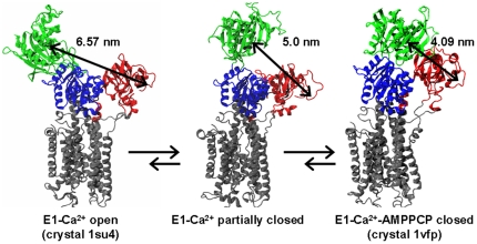
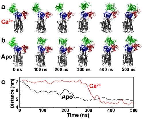
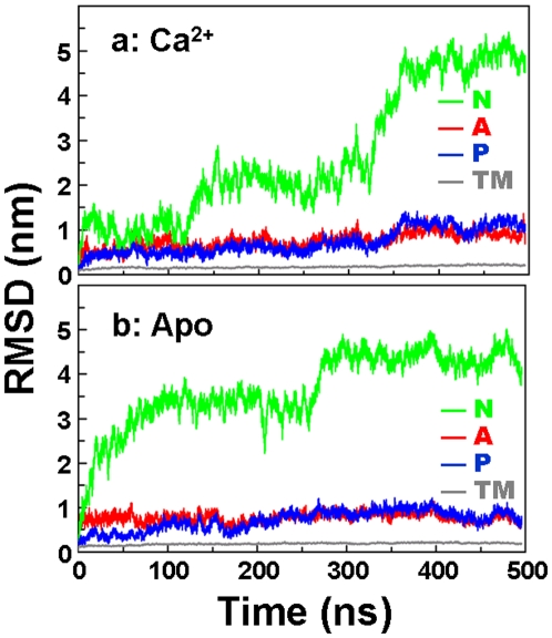
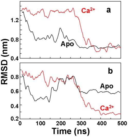
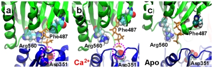
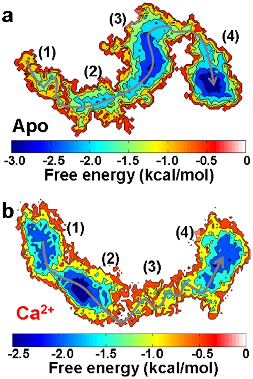
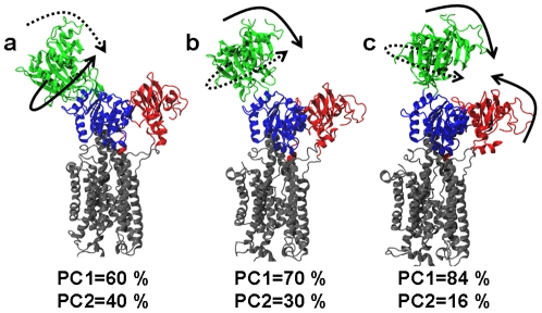
We have performed molecular dynamics (MD) simulations to elucidate, in atomic detail, the mechanism by which the sarcoplasmic reticulum Ca(2+)-ATPase (SERCA) is activated by Ca(2+). Crystal structures suggest that activation of SERCA occurs when the cytoplasmic head-piece, in an open (E1) conformation stabilized by Ca(2+), undergoes a large-scale open-to-closed (E1 to E2) transition that is induced by ATP binding. However, spectroscopic measurements in solution suggest that these structural states (E1 and E2) are not tightly coupled to biochemical states (defined by bound ligands); the closed E2 state predominates even in the absence of ATP, in both the presence and absence of Ca(2+). How is this loose coupling consistent with the high efficiency of energy transduction in the Ca(2+)-ATPase? To provide insight into this question, we performed long (500 ns) all-atom MD simulations starting from the open crystal structure, including a lipid bilayer and water. In both the presence and absence of Ca(2+), we observed a large-scale open-to-closed conformational transition within 400 ns, supporting the weak coupling between structural and biochemical states. However, upon closer inspection, it is clear that Ca(2+) is necessary and sufficient for SERCA to reach the precise geometrical arrangement necessary for activation of ATP hydrolysis. Contrary to suggestions from crystal structures, but in agreement with solution spectroscopy, the presence of ATP is not required for this activating transition. Principal component analysis showed that Ca(2+) reshapes the free energy landscape of SERCA to create a path between the open conformation and the activated closed conformation. Thus the malleability of the free energy landscape is essential for SERCA efficiency, ensuring that ATP hydrolysis is tightly coupled to Ca(2+) transport. These results demonstrate the importance of real-time dynamics in the formation of catalytically competent conformations of SERCA, with broad implications for understanding enzymatic catalysis in atomic detail.


Conflict of interest statement

Competing Interests: The authors have declared that no competing interests exist.

Figures

Figure 1
Figure 1. Proposed model of SERCA conformational dynamics induced by calcium.
The model is based on FRET experiments (Winters et al. [16]) showing that, in the presence of calcium, the SERCA headpiece samples a broad spectrum of conformations ranging from an open (E1-Ca2+ open, PDB:1su4) to a fully closed one (E1-Ca2+-AMPPCP closed, PDB:1vfp). SERCA is colored according to its four functional domains: N domain (green), P domain (blue), A domain (red) and TM domain (grey). We highlight the Cα-Cα distance between residues Met1 (A domain) and Lys515 (N domain), which were used as labeling sites to evaluate N-A interdomain distances.
Figure 2
Figure 2. Time-dependent conformational transitions of SERCA.
Structural snapshots from the simulations of Ca2+-bound (a) and Apo (b) SERCA, showing a complete transition from the open conformation (green N domain and red A domain far apart at 0 ns) to a closed one within 400 ns. (c) Cα-Cα distance between residues Met1 (A domain) and Lys515 (N domain) of Apo (black) and Ca2+-bound (red) SERCA.
Figure 3
Figure 3. Backbone RMSD of each functional domain of SERCA.
Domains are color-coded as indicated, as in Fig. 1. Backbone alignment used the TM domain as a reference.
Figure 4
Figure 4. Geometrical rearrangement of the cytosolic headpiece of SERCA.
(a) RMSD of the entire headpiece of Ca2+-bound (red) and Apo (black) SERCA; (b) RMSD calculated for the N and P domains only in the trajectories of Ca2+-bound (red) and Apo (black) SERCA. RMSD was calculated by superimposing the backbone atoms of domains N, P and A or N and P, respectively, onto the closed crystal structure of SERCA bound to Ca2+ and AMPPCP (PDB: 1vfp). Low RMSD values indicate similarity to the crystal structure.
Figure 5
Figure 5. Active site geometry of SERCA in atomic detail.
(a) Crystal structure of SERCA bound to calcium and AMPPCP (orange sticks). Simulations at 500 ns of (b) Ca2+-bound and (c) Apo SERCA. In both cases, the predicted orientation of AMPPCP was obtained by using rigid-body docking simulations. Residues that are necessary for nucleotide binding (Phe487 and Arg560) and autophosphorylation (Asp351) are shown as van der Waals spheres. The N and P domains are rendered as green and blue ribbons, respectively. The dashed magenta ovals highlight the proximity between the γ-phosphate of AMPPCP and the carboxyl group of Asp351 in a and b, but not in c.
Figure 6
Figure 6. PCA-based free energy landscape of the open-to-closed conformational transition of SERCA.
(a) Apo SERCA. (b) Ca2+-bound SERCA. The x and y axes correspond to the the first and second principal components, respectively. The gray arrow shows the preferred direction along the open-to-closed transition in the trajectories. Relevant regions discussed in the text are indicated by numbers in parentheses.
Figure 7
Figure 7. Directionality of motions along the open-to-closed transition of Ca2+-bound SERCA.
The motions correspond to (a) region 1, (b) region 2 and (c) region 3 of the energy landscape (Fig. 6B). Dashed and solid arrows represent motions described by the first and second principal components (PC1 and PC2), respectively. The percentage of contribution of each principal component (PC) that belongs to the essential space is shown below each structure.

References

    1. Henzler-Wildman K, Kern D. Dynamic personalities of proteins. Nature. 2007;450:964–72. - PubMed
    1. Frauenfelder H, McMahon B. Dynamics and function of proteins: the search for general concepts. Proc Natl Acad Sci U S A. 1998;95:4795–7. - PMC - PubMed
    1. Lange OF, Lakomek NA, Fares C, Schroder GF, Walter KF, et al. Recognition dynamics up to microseconds revealed from an RDC-derived ubiquitin ensemble in solution. Science. 2008;320:1471–5. - PubMed
    1. Bakan A, Bahar I. The intrinsic dynamics of enzymes plays a dominant role in determining the structural changes induced upon inhibitor binding. Proc Natl Acad Sci U S A. 2009;106:14349–54. - PMC - PubMed
    1. Boehr DD, Nussinov R, Wright PE. The role of dynamic conformational ensembles in biomolecular recognition. Nat Chem Biol. 2009;5:789–96. - PMC - PubMed

Publication types

MeSH terms

Substances