. 2011:2011:583439.
doi: 10.1155/2011/583439. Epub 2011 Aug 8.

Multiple myeloma: a review of imaging features and radiological techniques

Affiliations

Multiple myeloma: a review of imaging features and radiological techniques

C F Healy et al. Bone Marrow Res. 2011.

Abstract

The recently updated Durie/Salmon PLUS staging system published in 2006 highlights the many advances that have been made in the imaging of multiple myeloma, a common malignancy of plasma cells. In this article, we shall focus primarily on the more sensitive and specific whole-body imaging techniques, including whole-body computed tomography, whole-body magnetic resonance imaging, and positron emission computed tomography. We shall also discuss new and emerging imaging techniques and future developments in the radiological assessment of multiple myeloma.


Figures

Figure 1
Figure 1
Lateral radiograph skull: diffuse lytic lesions giving classical “pepper pot skull” appearance.
Figure 2
Figure 2
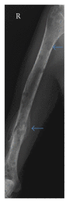
A-P radiograph right humerus: diffuse lytic lesions of the right humerus (arrowed) with old pathological fracture distal diaphysis (arrow).
Figure 3
Figure 3
Axial CT pelvis: diffuse myeloma involving the sacrum and iliac bones bilaterally, with cortical destruction of the left iliac bone (arrow).
Figure 4
Figure 4
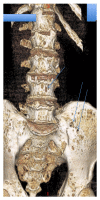
Volume rendering 3-dimensional reconstruction of lumbar spine and pelvis: multiple “punched-out” lytic lesions throughout lumbar spine and pelvis (arrow).
Figure 5
Figure 5
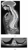
Low dose sagittal whole-body CT (a): note lytic lesion posterior aspect of T10 vertebral body (arrow). Background of extensive osseous permeation from myeloma. Axial CT thorax in the same patient at the level of T10 (b) identifying lytic infiltration of vertebral body (arrow).
Figure 6
Figure 6
Whole-body MRI coronal and selective axial STIR sequence. Left image: coronal STIR sequence demonstrating T2 bright myelomatous disease throughout the thoracic spine (rectangle). Centre image: coronal T1-weighted sequence demonstrating low signal marrow throughout the lumbar spine due the myelomatous infiltration (rectangle). Right image: 3 axial MRI images at the level of the vocal cords, lumbar spine, and ischium, T1-weighted sequence following administration of contrast.
Figure 7
Figure 7
(a) MRI sagittal T1-weighted sequence lumbar spine: diffuse permeative low signal myelomatous marrow lesions throughout the lumbar spine (arrow). (b) MRI sagittal T2-weighted STIR sequence (same patient): diffuse high signal myelomatous marrow lesions throughout the lumbar spine (arrow).
Figure 8
Figure 8
A-P radiograph right knee (a): 4 cm lucency medial femoral condyle (arrow), radiographically difficult to visualise. MRI coronal T2-weighted STIR sequence (b, same patient): high signal 4 cm plasmacytoma medial femoral condyle (arrow).
Figure 9
Figure 9
Axial fused PET/CT thorax at the level of the pulmonary bifurcation: massive right-sided chest wall plasmacytoma (arrow).
Figure 10
Figure 10
Axial CT thorax postintravenous contrast at the level of T6: diffuse bilateral hilar lymphadenopathy from biopsy-proven multiple myeloma (arrows).
Figure 11
Figure 11
Axial fused PET/CT at the level of T2 vertebra: extensive FDG-avid biopsy-proven amyloid left deltoid muscle in a patient with multiple myeloma (arrow).

References

    1. Landis SH, Murray T, Bolden S, Wingo PA. Cancer statistics, 1998. Ca-A Cancer Journal for Clinicians. 1998;48(1):6–29. - PubMed
    1. Desikan R, Barlogie B, Sawyer J, et al. Results of high-dose therapy for 1000 patients with multiple myeloma: durable complete remissions and superior survival in the absence of chromosome 13 abnormalities. Blood. 2000;95(12):4008–4010. - PubMed
    1. Waheed S, Shaughnessy JD, Van Rhee F, et al. International staging system and metaphase cytogenetic abnormalities in the era of gene expression profiling data in multiple myeloma treated with total therapy 2 and 3 protocols. Cancer. 2011;117(5):1001–1009. - PMC - PubMed
    1. Durie BGM, Salmon SE. A clinical staging system for multiple myeloma. Correlation of measured myeloma cell mass with presenting clinical features, response to treatment, and survival. Cancer. 1975;36(3):842–854. - PubMed
    1. Durie BGM. The role of anatomic and functional staging in myeloma: description of Durie/Salmon plus staging system. European Journal of Cancer. 2006;42(11):1539–1543. - PubMed

LinkOut - more resources