. 2011 Nov 2:11:321.
doi: 10.1186/1471-2148-11-321.

Structural and evolutionary divergence of eukaryotic protein kinases in Apicomplexa

Affiliations

Structural and evolutionary divergence of eukaryotic protein kinases in Apicomplexa

Eric Talevich et al. BMC Evol Biol. .

Abstract

Background: The Apicomplexa constitute an evolutionarily divergent phylum of protozoan pathogens responsible for widespread parasitic diseases such as malaria and toxoplasmosis. Many cellular functions in these medically important organisms are controlled by protein kinases, which have emerged as promising drug targets for parasitic diseases. However, an incomplete understanding of how apicomplexan kinases structurally and mechanistically differ from their host counterparts has hindered drug development efforts to target parasite kinases.

Results: We used the wealth of sequence data recently made available for 15 apicomplexan species to identify the kinome of each species and quantify the evolutionary constraints imposed on each family of apicomplexan kinases. Our analysis revealed lineage-specific adaptations in selected families, namely cyclin-dependent kinase (CDK), calcium-dependent protein kinase (CDPK) and CLK/LAMMER, which have been identified as important in the pathogenesis of these organisms. Bayesian analysis of selective constraints imposed on these families identified the sequence and structural features that most distinguish apicomplexan protein kinases from their homologs in model organisms and other eukaryotes. In particular, in a subfamily of CDKs orthologous to Plasmodium falciparum crk-5, the activation loop contains a novel PTxC motif which is absent from all CDKs outside Apicomplexa. Our analysis also suggests a convergent mode of regulation in a subset of apicomplexan CDPKs and mammalian MAPKs involving a commonly conserved arginine in the αC helix. In all recognized apicomplexan CLKs, we find a set of co-conserved residues involved in substrate recognition and docking that are distinct from metazoan CLKs.

Conclusions: We pinpoint key conserved residues that can be predicted to mediate functional differences from eukaryotic homologs in three identified kinase families. We discuss the structural, functional and evolutionary implications of these lineage-specific variations and propose specific hypotheses for experimental investigation. The apicomplexan-specific kinase features reported in this study can be used in the design of selective kinase inhibitors.


Figures

Figure 1
Figure 1
Kinome group composition. Composition of protein kinase major groups and selected apicomplexan-specific families (FIKK and ROPK) in each of the surveyed genomes. The schematic species tree along the left edge is constructed from published sources [36,92-94], and includes three outgroup kinomes for comparison: the dinoflagellate Perkinsus marinus, the diatom Thalassiosira pseudonana, and the yeast Saccharomyces cerevisiae. In the stacked bar chart associated with each genome, block width indicates number of genes found belonging to each major group of eukaryotic protein kinases; total bar width indicates total kinome size.
Figure 2
Figure 2
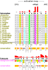
CHAIN alignment of the CDK subfamily activation loop. CHAIN alignment of the activation loop in the Pfcrk-5-like CDK subfamily ("Foreground") compared to the corresponding region in a large set of diverse eukaryotic CDK sequences ("Background"). The kinase-conserved DFG and APE motifs bordering the activation loop are indicated at the top, along with the subfamily-conserved PTxC motif. An asterisk indicates the position of the threonine observed to be phosphorylated in other CDKs, conserved in both the foreground and background. The histogram above each sequence set represents the differential levels of conservation between the two sets at each position, using logarithmic scaling. Dots above each alignment column indicate the contrasting conservation pattern determined by CHAIN. Note that the Apicomplexa (foreground, top) and Eukaryota (background, bottom) sets have different conservation patterns. In the sequence alignment itself, columns of the conserved pattern are colored according to the consensus residue type. The consensus residue types are listed below the alignment. Weighted residue frequencies are shown in the following rows, in units of integer tenths (e.g. "9" indicates conservation of 90-100%). The number of sequences in each set are shown in parentheses. A complete CHAIN alignment of these sequences is provided in Additional File 4.
Figure 3
Figure 3
Logo of the CDK subfamily cyclin-binding motif. Logo of the aligned activation loop sequences in members of the Pfcrk-5-like CDK subfamily, generated by WebLogo [95]. Letter height represents information content; large letters indicated residues conserved within the subfamily.
Figure 4
Figure 4
CDPK subfamily roles of αC helix arginine and activation-loop threonine. Structures of several different CDPKs in C. parvum, demonstrating several proposed interactions for the αC helix arginine distinctive of an alveolate-specific CDPK subfamily. (A) A member of the background set of CDPKs [PDB:3DFA] has a threonine (T50), shown in cyan, in position to form a hydrogen bond with an aspartate (D47), gray, which caps the αC helix. This threonine corresponds to the subfamily-conserved arginine; however, the threonine here is not conserved in the background set of CDPKs. (B) In a structure of a member of the CDPK subfamily [PDB:2QG5], the subfamily-conserved arginine (R69, cyan) appears similarly positioned to interact with the aspartate (D66, blue) at the end of the αC helix, potentially stabilizing the cap. (C) Chain A of the same structure shows the distinctive arginine oriented inward, capable of hydrogen-bonding with the kinase-conserved DFG motif (side chains colored magenta). (D) In another structure of the same CDPK-subfamily protein [PDB:2QG5], the arginine is positioned toward a subfamily-conserved threonine in the activation loop (T184), shown in cyan. The distance between the R69 and T184 side chains is 6Å, which could accomodate a phosphate group attached to the threonine and a hydrogen bond between the phosphothreonine and the arginine.
Figure 5
Figure 5
CLK docking site. Three contrastingly conserved residues involved in substrate recognition and docking in human Clk2 [PDB:3NR9] and the P. falciparum CLK, PfLAMMER [PDB:3LLT]. (A) Global view of the docking site, illustrating the position of the substrate RS domain and phosphorylation site. The contrastingly conserved resides are shown in cyan. (B) Human Clk2. A trio of constrastingly conserved residues (cyan), along with a nearby phenylalanine (gray), form a network of hydrogen bonds. The conserved histidine (H346) is positioned to interact with the substrate P-2 position. (C) In PfLAMMER, the three residues (cyan) are conserved as different types. A glutamine (Q739) replaces the histidine in human Clk2 seen to interact with the substrate P-2 position. The hydrogen bonding network is different: A leucine (L772) replaces the threonine seen in Clk2; an arginine (R775), corresponding to a glutamate in Clk2, is directed away from the other two conserved residues; and the glutamine (Q739) instead forms a hydrogen bond with a nearby threonine.
Figure 6
Figure 6
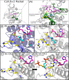
CLK coordination of substrate-binding and catalytic regions. Interactions between key residues in the substrate-binding region and the catalytic HTD motif are mediated by conserved residues in the activation loop. (A) Structural context of features in PfLAMMER [PDB:3LLT], showing the activation loop in green and the catalytic loop in magenta. Conserved residues are displayed in "sticks" representation. A contrastingly conserved asparagine, distinctive of chromalveolate CLKs, is indicated in cyan, and three other residues conserved throughout the CLK family are shown in yellow. (B) In PfLAMMER, the distinctive asparagine (N736) forms hydrogen bonds with the CMGC-conserved arginine (R741), the backbone of the alanine in the APE motif, the backbone of the threonine in the catalytic HTD motif, and, mediated by a water molecule, a subfamily-conserved serine in the αF helix. (C) In human SRPK1, several of the hydrogen bonds formed by the glutamine Q513 are analogous to those formed by the N736 in apicomplexans. (D) and (E) Two structures of human Clk1. In the unphosphorylated structure [PDB:1Z57], left, the serine corresponding to PfLAMMER N736 (S341) and the adjacent CLK-conserved threonine (T342) are oriented in an "in" conformation, interacting with the catalytic motif (HTD) but not with the conserved arginines (R343, R346). In the phosphorylated structure [PDB:2VAG], right, the serine (pS341) and threonine (pT342) are flipped to an "out" conformation, breaking the interaction with the catalytic motif. One arginine (R343) moves to occupy the area vacated by the phosphorylated serine S341, while the other (R346) now interacts with the backbone of the phosphorylated serine. Phosphates are shown in orange. Images of PDB structures were rendered using PyMOL [69].
Figure 7
Figure 7
CLK β7-β8 hairpin insert and anchoring residues. Comparison of the residue interactions anchoring the β-hairpin insert to the kinase C-lobe in solved structures of PfLAMMER [PDB:3LLT] and human Clk2 [PDB:3NR9]. (A) Both structures superimposed, with corresponding key residues shown in "sticks" representation. The contrastingly conserved residue (from CHAIN analysis) is highlighted cyan: D653 in PfLAMMER, Q266 in human Clk2. A residue of interest near the base of the hairpin insert, discussed in the text, is shown in yellow; its type is not strongly conserved within apicomplexan CLKs. Two residues in the loop of the hairpin, colored green, are inserts in PfLAMMER relative to Clk2; they appear anchored to the kinase C-lobe by interactions with a lysine, dark blue. (B) Human Clk2, showing side chains near the residues of interest. A hydrogen bond appears between the αE-helix glutamine (cyan) and the backbone of a valine (yellow) near the base of the hairpin insert. (C) In PfLAMMER, the two residues of interest, D653 (cyan) and T711 (yellow), do not interact directly; each instead forms several novel hydrogen bonds with other nearby residues, shown in green and blue, corresponding to those shown in green and gray in the human Clk2 structure.

References

    1. Roos DS. Genetics. Themes and variations in apicomplexan parasite biology. Science. 2005;309(5731):72–3. - PubMed
    1. Renslo AR, McKerrow JH. Drug discovery and development for neglected parasitic diseases. Nature chemical biology. 2006;2(12):701–10. - PubMed
    1. Sibley LD. Intracellular parasite invasion strategies. Science. 2004;304(5668):248–53. - PubMed
    1. Hammarton T. The cell cycle of parasitic protozoa: potential for chemotherapeutic exploitation. Progress In Cell Cycle Research. 2003;5:91–101. - PubMed
    1. Doerig C, Abdi A, Bland N, Eschenlauer S, Dorin-Semblat D, Fennell C, Halbert J, Holland Z, Nivez MP, Semblat JP, Sicard A, Reininger L. Malaria: targeting parasite and host cell kinomes. Biochimica et biophysica acta. 2010;1804(3):604–12. - PubMed

Publication types

Substances

LinkOut - more resources