Clinical Trial
. 2011 Nov 3;365(18):1673-83.
doi: 10.1056/NEJMoa1106152.

Inducible apoptosis as a safety switch for adoptive cell therapy

Affiliations
Clinical Trial

Inducible apoptosis as a safety switch for adoptive cell therapy

Antonio Di Stasi et al. N Engl J Med. .

Abstract

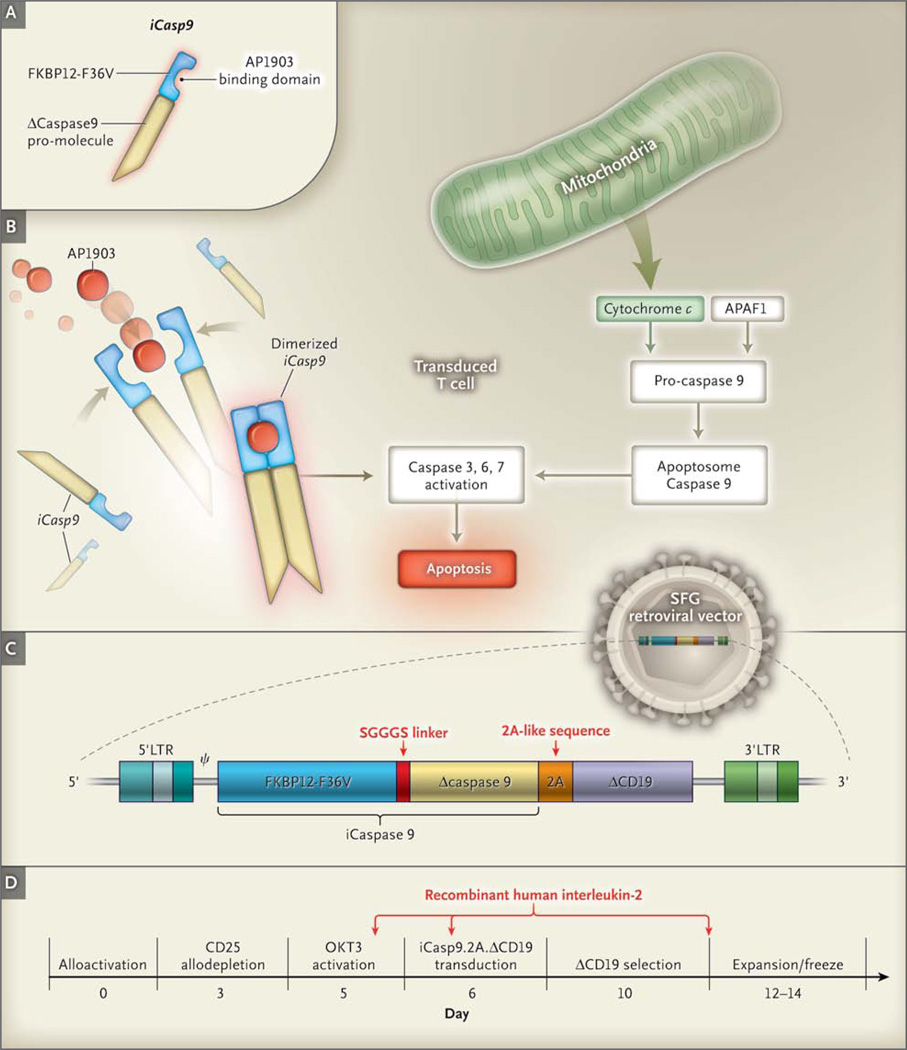
Background: Cellular therapies could play a role in cancer treatment and regenerative medicine if it were possible to quickly eliminate the infused cells in case of adverse events. We devised an inducible T-cell safety switch that is based on the fusion of human caspase 9 to a modified human FK-binding protein, allowing conditional dimerization. When exposed to a synthetic dimerizing drug, the inducible caspase 9 (iCasp9) becomes activated and leads to the rapid death of cells expressing this construct.

Methods: We tested the activity of our safety switch by introducing the gene into donor T cells given to enhance immune reconstitution in recipients of haploidentical stem-cell transplants. Patients received AP1903, an otherwise bioinert small-molecule dimerizing drug, if graft-versus-host disease (GVHD) developed. We measured the effects of AP1903 on GVHD and on the function and persistence of the cells containing the iCasp9 safety switch.

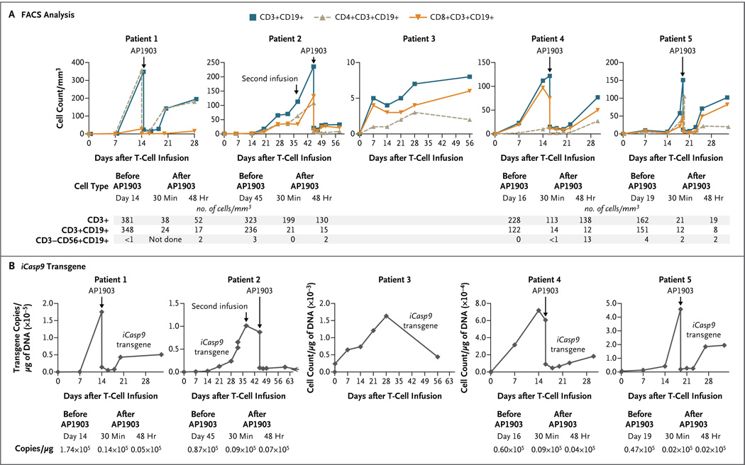
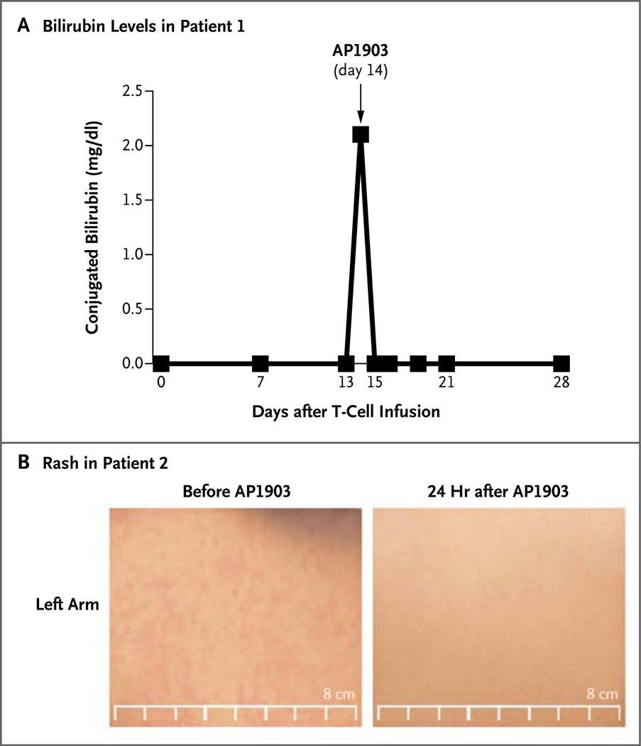
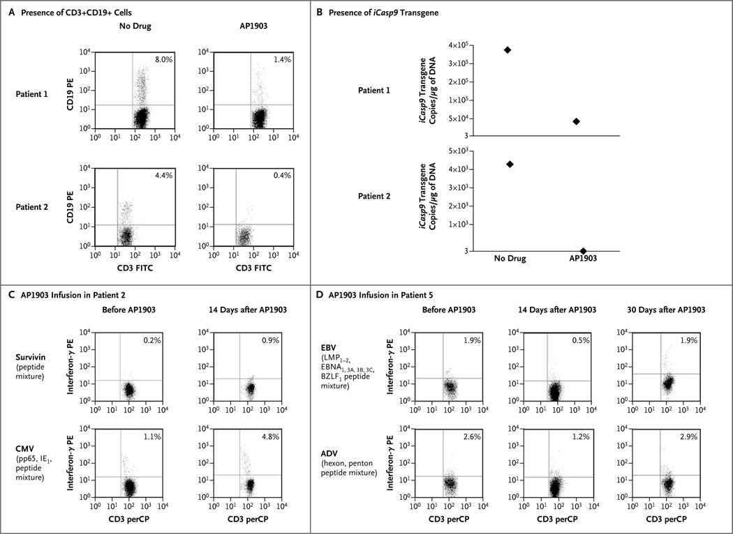
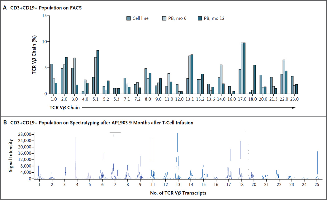
Results: Five patients between the ages of 3 and 17 years who had undergone stem-cell transplantation for relapsed acute leukemia were treated with the genetically modified T cells. The cells were detected in peripheral blood from all five patients and increased in number over time, despite their constitutive transgene expression. A single dose of dimerizing drug, given to four patients in whom GVHD developed, eliminated more than 90% of the modified T cells within 30 minutes after administration and ended the GVHD without recurrence.

Conclusions: The iCasp9 cell-suicide system may increase the safety of cellular therapies and expand their clinical applications. (Funded by the National Heart, Lung, and Blood Institute and the National Cancer Institute; ClinicalTrials.gov number, NCT00710892.).


Figures

Figure 1
Figure 1. Generation of Transgene and Function of Activated iCasp9
In Panel A, the suicide gene iCasp9 is shown to consist of the sequence of the human FK506-binding protein, FKBP12, with an F36V mutation, connected through a series of amino acids to the gene encoding human caspase 9. FKBP12-F36V binds with high affinity to a small-molecule dimerizing agent, AP1903. Panel B shows that physiological activation of the intrinsic apoptosis pathway requires a cytoplasmic protein, apoptotic peptidase activating factor 1 (APAF1), which binds with cytochrome c to form an oligomeric apoptosome, which in turn binds and cleaves caspase 9 preproprotein, releasing an activated form of the peptidase and resulting in a caspase cascade that ends in apoptosis. In transduced cells, the administration of AP1903 leads to dimerization of iCasp9, thereby bypassing activation of the initial mitochondrial apoptotic pathway. Panel C shows the structure of the iCaspase9.2A.ΔCD19 bicistronic transgene (i.e., one with two cistrons, the loci responsible for generating a protein), comprising the iCasp9 sequence, with truncated CD19 (ΔCD19) serving as the selectable marker. The sequence cassette is then incorporated into the SFG retroviral vector. Panel D shows the manufacturing process, along with the day of each manipulation. The arrows indicate the times of adding recombinant human interleukin-2 to the cultures. LTR denotes long terminal repeat, OKT3 mouse antihuman CD3 monoclonal antibody, and SGGGS Ser-Gly-Gly-Gly-Ser.
Figure 2
Figure 2. Detection of iCasp9-Transduced T Cells in Peripheral Blood
Panel A shows fluorescence-activated cell sorting (FACS) analysis for iCasp9-transduced T cells (CD3+, CD3+CD19+, CD4+CD19+, CD8+CD19+, or CD3−CD56+CD19+, with the cell count given per cubic millimeter of peripheral blood) from five patients who received cellular therapy after undergoing HLA-haploidentical stem-cell transplantation for relapsed leukemia. Skin or liver graft-versus-host disease (GVHD) developed in Patients 1, 2, 4, and 5, who had high levels of engraftment. Each of these patients received a single dose of AP1903. The tables beneath the graphs show the cell counts before and after treatment with AP1903. Panel B shows the number of copies of the iCasp9 transgene per microgram of DNA obtained from peripheral-blood mononuclear cells, evaluated by means of real-time quantitative polymerase-chain-reaction (PCR) amplification, for each patient at time points corresponding to those in Panel A before and after AP1903 infusion. The tables beneath the graphs show the actual quantitative PCR values before and after treatment, revealing a diminution by a factor of 100 (2 log) in the signal from the iCasp9 transgenic cells.
Figure 3
Figure 3. Rapid Reversal of GVHD after Treatment with AP1903
Panel A shows the normalization of bilirubin levels in Patient 1 within 24 hours after treatment with AP1903. To convert the values for bilirubin to micromoles per liter, multiply by 17.1. Panel B shows the disappearance of rash from the left arm of Patient 2 within 24 hours after treatment.
Figure 4
Figure 4. Persistence of Drug Sensitivity and Antiviral Function of CD3+CD19+ T Cells after Treatment with AP1903 in Vivo
Panel A shows the presence of CD3+CD19+ cells and the iCasp9 transgene in Patients 1 and 2. Cells were counted and incubated with antibodies labeled with fluorochrome (phycoerythrin [PE] or fluorescein isothiocyanate [FITC]) targeting CD19+ cells (PE) and CD3+ cells (FITC). CD3+CD19+ T cells remained within the CD3+ population in the peripheral blood after treatment with AP1903 for 5 months in Patient 1 and for 9 months in Patient 2. These CD3+CD19+ cells retained sensitivity to AP1903 in vitro, as assessed both by the reduction in the number of CD3+CD19+ cells on fluorescence-activated cell sorting (FACS) analysis (as measured per microliter of peripheral blood) (Panel A) and by quantitative polymerase-chain-reaction (PCR) analysis of the iCasp9 gene before and after exposure to the dimerizing drug (Panel B). As shown in Panel A, values for Patient 1 were 2290 lymphocytes per cubic millimeter with a CD3+ count of 41%, and values for Patient 2 were 1380 lymphocytes per cubic millimeter with a CD3+ count of 35%. As indicated on the y axis in Panel B, 3 is the minimum number of copies of iCasp9 that can be detected on quantitative PCR. CD3+CD19+ gene-modified T cells that were collected from Patient 2 (Panel C) and Patient 5 (Panel D) were responsive to viral peptide mixtures and survivin peptide mixture (in Patient 2) before the administration of AP1903, as shown by the presence of interferon-γ–positive CD3+CD19+ T cells in the peptide-stimulated cultures. The assessment of the recovering CD3+CD19+ population at 14 days in Patient 2 and at 14 and 30 days in Patient 5 after AP1903 infusion to treat GVHD showed the persistence of these antigen-specific cells in the absence of recurrent GVHD. ADV denotes adenovirus, BZLF1 BamHI Z leftward reading frame, EBNA Epstein–Barr nuclear antigen, IE1 immediate early protein, LMP latent membrane protein, perCP peridinin chlorophyll protein, and pp65 phosphoprotein 65.
Figure 5
Figure 5. Polyclonality of Recovering T Cells in Patient 1
The analysis of the repertoire of T-cell receptor variable beta chain (TCR Vβ) in the CD3+CD19+ selected population, which recovered 6 months after the administration of the dimerizing drug in Patient 1, shows a polyclonal pattern of expression, as measured by reactivity to monoclonal antibodies directed against TCR Vβ chain families and measured on FACS analysis (Panel A), as well as by multiplex polymerase-chain-reaction–based spectratyping to analyze the number of distinct TCR Vβ transcripts present (Panel B). PB denotes peripheral blood.

Comment in

  • Eliminating cells gone astray.
    Sadelain M. Sadelain M. N Engl J Med. 2011 Nov 3;365(18):1735-7. doi: 10.1056/NEJMe1109971. N Engl J Med. 2011. PMID: 22047566 No abstract available.

References

    1. Daley GQ, Scadden DT. Prospects stem cell-based therapy. Cell. 2008;132:544–548. - PubMed
    1. Williams DA, Keating A. Enhancing research in regenerative medicine. Blood. 2010;116:866–867. - PubMed
    1. Appelbaum FR. Haematopoietic cell transplantation as immunotherapy. Nature. 2001;411:385–389. - PubMed
    1. Kolb HJ. Graft-versus-leukemia effects of transplantation and donor lymphocytes. Blood. 2008;112:4371–4383. - PubMed
    1. Boztug K, Schmidt M, Schwarzer A, et al. Stem-cell gene therapy for the Wiskott–Aldrich syndrome. N Engl J Med. 2010;363:1918–1927. - PMC - PubMed

Publication types

MeSH terms

Associated data