Review
. 2012 Feb;8(1 Suppl):S1-68.
doi: 10.1016/j.jalz.2011.09.172. Epub 2011 Nov 2.

The Alzheimer's Disease Neuroimaging Initiative: a review of papers published since its inception

Affiliations
Review

The Alzheimer's Disease Neuroimaging Initiative: a review of papers published since its inception

Michael W Weiner et al. Alzheimers Dement. 2012 Feb.

Abstract

The Alzheimer's Disease Neuroimaging Initiative (ADNI) is an ongoing, longitudinal, multicenter study designed to develop clinical, imaging, genetic, and biochemical biomarkers for the early detection and tracking of Alzheimer's disease (AD). The study aimed to enroll 400 subjects with early mild cognitive impairment (MCI), 200 subjects with early AD, and 200 normal control subjects; $67 million funding was provided by both the public and private sectors, including the National Institute on Aging, 13 pharmaceutical companies, and 2 foundations that provided support through the Foundation for the National Institutes of Health. This article reviews all papers published since the inception of the initiative and summarizes the results as of February 2011. The major accomplishments of ADNI have been as follows: (1) the development of standardized methods for clinical tests, magnetic resonance imaging (MRI), positron emission tomography (PET), and cerebrospinal fluid (CSF) biomarkers in a multicenter setting; (2) elucidation of the patterns and rates of change of imaging and CSF biomarker measurements in control subjects, MCI patients, and AD patients. CSF biomarkers are consistent with disease trajectories predicted by β-amyloid cascade (Hardy, J Alzheimers Dis 2006;9(Suppl 3):151-3) and tau-mediated neurodegeneration hypotheses for AD, whereas brain atrophy and hypometabolism levels show predicted patterns but exhibit differing rates of change depending on region and disease severity; (3) the assessment of alternative methods of diagnostic categorization. Currently, the best classifiers combine optimum features from multiple modalities, including MRI, [(18)F]-fluorodeoxyglucose-PET, CSF biomarkers, and clinical tests; (4) the development of methods for the early detection of AD. CSF biomarkers, β-amyloid 42 and tau, as well as amyloid PET may reflect the earliest steps in AD pathology in mildly symptomatic or even nonsymptomatic subjects, and are leading candidates for the detection of AD in its preclinical stages; (5) the improvement of clinical trial efficiency through the identification of subjects most likely to undergo imminent future clinical decline and the use of more sensitive outcome measures to reduce sample sizes. Baseline cognitive and/or MRI measures generally predicted future decline better than other modalities, whereas MRI measures of change were shown to be the most efficient outcome measures; (6) the confirmation of the AD risk loci CLU, CR1, and PICALM and the identification of novel candidate risk loci; (7) worldwide impact through the establishment of ADNI-like programs in Europe, Asia, and Australia; (8) understanding the biology and pathobiology of normal aging, MCI, and AD through integration of ADNI biomarker data with clinical data from ADNI to stimulate research that will resolve controversies about competing hypotheses on the etiopathogenesis of AD, thereby advancing efforts to find disease-modifying drugs for AD; and (9) the establishment of infrastructure to allow sharing of all raw and processed data without embargo to interested scientific investigators throughout the world. The ADNI study was extended by a 2-year Grand Opportunities grant in 2009 and a renewal of ADNI (ADNI-2) in October 2010 through to 2016, with enrollment of an additional 550 participants.


Figures

Figure 1
Figure 1. Generation of soluble β-amyloid fragments from amyloid precursor protein
From [7].
Figure 2
Figure 2. Model for AD disease progression
From [14].
Figure 3
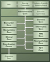
Figure 3. ADNI structure and organization
Figure 4
Figure 4. Roles of biomarkers in AD drug development
ADMET- absorption, distribution, metabolism, excretion, toxicity; BBB= blood-brain barrier; POP = proof of principle. From [37].
Figure 5
Figure 5. AD drug development
Black arrows show the phases of drug development; the brick-colored arrows show the ADNI biomarkers that could be used in that stage. From [37].
Figure 6
Figure 6. Steps of multi-atlas segmentation
(I) non-rigid registration used to register all atlases to patient data, (II) classifier fusion using majority voting for producing class labels form all voxels, and (III) post-processing of multi-atlas segmentation result by various algorithms taking into account intensity distributions of different structures. From [61].
Figure 7
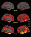
Figure 7. Group differences in average thickness (mm) for left hemisphere
Top row: NC vs. MCI non-converters (SMCI); middle row: NC vs. MCI converters (MMCI); bottom row: NC vs. AD. Left mesial views, right lateral views. The scale ranges from <20.3 (yellow) to >10.3 (cyan) mm thickness. Areas on the red-yellow spectrum indicate regions of thinning with disease: approximate color scale in mm is 20.05 to 20.15 dark red, 20.20 bright red, 20.25 orange, and <20.30 yellow. For thicker regions: 10.05 to 10.15 blue. Any differences smaller than 60.05 mm are gray. From [109].
Figure 8
Figure 8. Annual atrophy rates as a function of degree of clinical impairment
Clinical impairment measured using baseline CDR-SB scores. Mean atrophy rates are represented as a percent change in neocortical volume and mapped onto the lateral (left), ventral (middle), and medial (right) pial surface of the left hemisphere. These data demonstrate that atrophy rates are most prominent in posterior brain regions early in the course of disease, spreading to anterior regions as the level of impairment increases, with relative sparing of sensorimotor regions. From [111].
Figure 9
Figure 9. Distribution of atrophy scores used to classify subjects with MCI
MCI atrophy score was derived from LDA trained on data from all control subjects and subjects with AD. Discriminant model assumed equal prior group probabilities. Individuals were classified as having control phenotype if their scores were above −0.33. Cutoff score was chosen to maximize overall accuracy of classifying control subjects and subjects with AD on whom this model was trained. Average atrophy score for subjects with MCI was −0.50. Atrophy score is not normally distributed (Kolmogorov-Smirnov test = 0.73, df= 175, P = .025) but shows evidence of bimodal distribution. From [117].
Figure 10
Figure 10. Individual trajectories of hippocampal volume change
Thick black lines indicate the mean trajectory change of each group. From [121].
Figure 11
Figure 11. APOE gene effects on regional brain volumes
Maps show the mean percent differences in regional brain volumes for four different group comparisons. Percent differences are displayed on models of the regions implicated: (a) ventricular CSF, (b) sulcal CSF, (c) hippocampi, and (d) temporal lobes; dotted lines show the boundary of the hippocampus. From [112].
Figure 12
Figure 12. Group differences in regional shape deformations
Am - amygdala, Hp - hippocampus, V - ventricles, iLV - inferior lateral ventricles, Cd - caudate, Pu - putamen, Pa - globus pallidus, Th - thalamus. From [122].
Figure 13
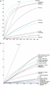
Figure 13. CDF plots for voxel-wise correlation of progressive temporal lobe tissue loss in MCI, AD, and pooled groups
(a) Correlations with various biomarker indices including Aβ42 (AB142), tau protein (TAU), phosphorylated-tau 181 (PTAU), tau/Aβ42 ratio (TAUAB), and ptau/Aβ42 ratio (PTAUAB), and (b) correlations with various clinical measures. From [113].
Figure 14
Figure 14. Association of regional brain tissue volumes with BMI
These represent the estimated degree of tissue excess or deficit at each voxel, as a percentage, for every unit increase in BMI, after statistically controlling for the effects age, sex, and education on brain structure. Images are in radiological convention (left side of the brain shown on the right) and are displayed on a specially constructed average brain template created from the subjects within each cohort (mean deformation template, or MDT). From [134].
Figure 15
Figure 15. Correlations between biomarker levels, structural abnormalities and cognitive performance in APOE ε4 carriers and non-carriers
Smoothed biomarker (A and B) or STAND (C) z-score curves plotted as a function of cognitive performance (MMSE). STAND = Structural Abnormality Index. From [128].
Figure 16
Figure 16. The episodic memory network
Along with the hippocampal formation, the cortical areas shown here are part of the episodic memory network. Shown here are pial cortical representations of selected parcellations in the left hemisphere. From left to right: medial, ventral and lateral views. From [226].
Figure 17
Figure 17. Biomarker trajectories through disease progression
For each biomarker, individual Z scores are plotted against ADAS-Cog scores, and the fitted sigmoid curve is displayed. Full circles denote healthy controls, full squares MCI patients converted to AD, empty circles early AD, and full triangles late AD patients. Sigmoid fitting was better than linear fitting for tau, Aβ42 and hippocampus (for the latter: sigmoid non-significantly better than linear); linear fitting was better for FDG-PET. From [154].
Figure 18
Figure 18. Separation of control, MCI and AD patients using a CSF Aβ42/t-tau mixed model signature
A combined CSF Aβ42/t-tau mixed model was applied to the subject groups. Densities of each signature are represented with confidence ellipses, and signature membership of the subject based on the mixture is indicated with the corresponding color (signature 1 is the Alzheimer disease [AD] signature [red]; signature 2 is the healthy signature [green]). From [160].
Figure 19
Figure 19. Association between temporal lobe atrophy and conversion to AD
Subjects who converted from MCI to AD over a period of 1 year after their first scan were coded as “1”; non-converters were coded as “0”. A negative correlation suggests that temporal lobe degeneration predicts future conversion to AD. From [112].
Figure 20
Figure 20. Effect size of imaging biomarkers for MCI-converters vs. MCI-non-converters
Effect sizes (Cohen’s D) of the comparison between MCI-Stable (MCI non-converter) and MCI-Converter groups evaluated for selected imaging biomarkers. From [114].
Figure 21
Figure 21. Significance maps of correlation between ventricular shape and cognitive decline
Significance maps correlate baseline ventricular shape with subsequent decline, over the following year, in 3 commonly used clinical scores. From [126].
Figure 22
Figure 22. Maps of associations with MMSE scores at baseline and 1 year later, MCI-to-AD conversion, and CSF concentrations of tau
3D maps show areas of significant associations between local volumetric atrophy in the caudate and MMSE scores at baseline and after a 1-year follow-up interval, with p-values color-coded at each surface voxel. From [131].
Figure 23
Figure 23. PIB-PET and MRI comparisons of MCI-converter vs MCI- non-converters
Left: Mild cognitive impairment progressor, Top: positive PIB PET. Bottom: MRI illustrating atrophic hippocampi and ventricular enlargement. Right: Mild cognitive impairment non-progressor. Top: negative PIB PET with non-specific white matter retention but no cortical retention. Bottom: MRI illustrating normal hippocampi and no ventricular enlargement. From [153].
Figure 24
Figure 24. Mean biomarker levels (t-tau, p-tau and Aβ42) for the APOE genotype groups
The APOE 2 carriers are represented in black, the 3 homozygotes in grey and the 4 carriers in white. The CSF Aβ42 levels show a significant stepwise trend downward, from 2 carriers to 3 homozygotes to 4 carriers; whereas the t-tau and the p-tau levels show the opposite trend. From [209].
Figure 25
Figure 25. Worldwide ADNI sites
NA-ADNI, North American ADNI; Arg-ADNI, Argentinean ADNI; E-ADNI, European ADNI; C-ADNI, Chinese ADNI; K-ADNI, Korean ADNI; J-ADNI, Japanese ADNI; T-ADNI, Taiwanese ADNI; A-ADNI, Australian ADNI.

References

    1. Hardy J. Alzheimer’s disease: the amyloid cascade hypothesis: an update and reappraisal. J Alzheimers Dis. 2006;9(3 Suppl):151–153. - PubMed
    1. Mueller SG, Weiner MW, Thal LJ, Petersen RC, Jack C, Jagust W, et al. The Alzheimer’s disease neuroimaging initiative. Neuroimaging Clin N Am. 2005;15(4):869–877. xi–xii. - PMC - PubMed
    1. Weiner MW, Aisen PS, Jack CR, Jr, Jagust WJ, Trojanowski JQ, Shaw L, et al. The Alzheimer’s disease neuroimaging initiative: progress report and future plans. Alzheimers Dement. 2010;6(3):202–211. e7. - PMC - PubMed
    1. Frisoni GB, Weiner MW. Alzheimer’s Disease Neuroimaging Initiative special issue. Neurobiol Aging. 2010;31(8):1259–1262. - PubMed
    1. Petersen RC, Roberts RO, Knopman DS, Boeve BF, Geda YE, Ivnik RJ, et al. Mild cognitive impairment: ten years later. Arch Neurol. 2009;66(12):1447–1455. - PMC - PubMed