. 2011 Nov 2;480(7378):530-3.
doi: 10.1038/nature10639.

Adherens junction protein nectin-4 is the epithelial receptor for measles virus

Affiliations

Adherens junction protein nectin-4 is the epithelial receptor for measles virus

Michael D Mühlebach et al. Nature. .

Abstract

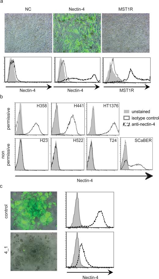
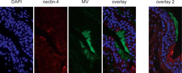
Measles virus is an aerosol-transmitted virus that affects more than 10 million children each year and accounts for approximately 120,000 deaths. Although it was long believed to replicate in the respiratory epithelium before disseminating, it was recently shown to infect initially macrophages and dendritic cells of the airways using signalling lymphocytic activation molecule family member 1 (SLAMF1; also called CD150) as a receptor. These cells then cross the respiratory epithelium and transport the infection to lymphatic organs where measles virus replicates vigorously. How and where the virus crosses back into the airways has remained unknown. On the basis of functional analyses of surface proteins preferentially expressed on virus-permissive human epithelial cell lines, here we identify nectin-4 (ref. 8; also called poliovirus-receptor-like-4 (PVRL4)) as a candidate host exit receptor. This adherens junction protein of the immunoglobulin superfamily interacts with the viral attachment protein with high affinity through its membrane-distal domain. Nectin-4 sustains measles virus entry and non-cytopathic lateral spread in well-differentiated primary human airway epithelial sheets infected basolaterally. It is downregulated in infected epithelial cells, including those of macaque tracheae. Although other viruses use receptors to enter hosts or transit through their epithelial barriers, we suggest that measles virus targets nectin-4 to emerge in the airways. Nectin-4 is a cellular marker of several types of cancer, which has implications for ongoing measles-virus-based clinical trials of oncolysis.


Figures

Figure 1
Figure 1. Identification of nectin-4 as candidate MV receptor
a, Nectin-4 mediates MV entry. CHO-K1 cells were not transfected (mock), or transfected with the expression plasmids nectin-4 or MST1R. Top row: after transfection cells were infected with a MV expressing GFP. Bottom row: expression of nectin-4 and MST1R (interrupted black lines), as documented by FACS analysis. Dark grey lines: isotype controls; light grey shadings: unstained controls. b, FACS analysis of nectin-4 expression levels in different cells. Cell lines H358, H441 and HT1376, top row, are permissive for MV infection. Cell lines H23, H522, T24, and SCaBER are non-permissive. Antibody LS-C37483 was used. c, siRNA-mediated knock-down of nectin-4 expression in H358 cells (left panels). Control (top row) or nectin-4 specific 4_1 siRNA (bottom row) were transfected. After transfections cells were infected with a MV expressing GFP. FACS analysis (right panels) indicated more than 90% reduction of nectin-4 expression on the cell surface.
Figure 2
Figure 2. V domain of nectin-4 supports strong binding to MV H
a and b, MV infection competition assays in H358 cells. For (a), N4VCC-Fc (grey bars) with all three immunoglobulin domains, and N4V-Fc (black bars) with only the V domain were used. As control soluble immunoglobulin superfamily protein CTLA4-Fc (cytotoxic T lymphocyte antigen 4, also named CD152) was used (white bars). For (b), antibodies N4.40, recognizing one of the C domains, or N4.61 recognizing the V domain, were used. All assays were done in triplicate (n=3); mean and standard deviation of the number of observed syncytia per well are indicated on the vertical axis. Concentration of the undiluted protein solution is indicated on the horizontal axis. For dimeric N4VCC-Fc 10 μg/ml are 71 nM; no, no protein added. c and d, non-reducing gel electrophoresis analysis of binding of soluble H and purified virus particles to different soluble receptors. Molecular weight markers are indicated. N4VCC-Fc and N4V-Fc are dimeric proteins of 140 kDa and 90 kDa respectively. The 260 kDa observed for N4VCC-Fc probably corresponds to association of two dimers through C domain dependent homophilic interactions. For (d), CTLA4-Fc loaded at the same concentration did not cross-react with soluble H (right top panel), but was detected by protein A-HRP with the same efficiency (right bottom panel). e, surface plasmon resonance analyses of H binding to SLAM. Multiple cycles of association and dissociation were performed using receptor concentrations ranging from 50 to 800 nM (color coding on top). Horizonal axis: time in seconds; vertical axis, absorbance units. f, surface plasmon resonance analyses of H binding to nectin-4.
Figure 3
Figure 3. Nectin-4 is necessary for MV infection of well-differentiated human airway epithelia
a, nectin-4 mRNA expression assessed by quantitative RT PCR and presented as fold change relative to Calu-3 cells. For human airway epithelial cells (hAEC), the bar represents data from 6 different donor samples (n=6). For cell lines (Calu-3, HEK293, Huh7) each bar represents 3 separate RNA preparations (n=3). b, knockdown of nectin-4 mRNA in hAEC. Levels of nectin-4 mRNA were assessed 72 hours after transfection of a control siRNA (left column) or a nectin-4 specific siRNA (right column) (n=6). c, lack of MV infection in hAEC after nectin-4 mRNA knockdown. GFP-positive infectious centers were counted 3 days following MV infection of hAEC transfected with a negative control siRNA (left column) or nectin-4 siRNA (n=9, nine epithelial preparations from a single donor lung). All data are expressed as mean +/- standard deviation. We note that if the well with 17 infectious centers is excluded, p improves to 0.0055. However, the infectious center average drops to 2.75 in the non-treated control.
Figure 4
Figure 4. Nectin-4 expression and infection of monkey tracheal epithelium
Microscopic analyses of tracheal tissue obtained from cynomolgus macaques 12 days after inoculation with MV. From left to right: nuclear staining by DAPI (blue); nectin-4 staining with a mixture of two antibodies (red); MV infection revealed through GFP expression (green); overlay of the three panels; overlay of three panels of another section (overlay 2). Frozen sections were photographed at 4000-fold magnification with oil immersion.

References

    1. Vaccines: the case of measles. Nature. 2011;473:434–435. doi:10.1038/473434a. - PubMed
    1. Chen SY, et al. Health care-associated measles outbreak in the united states after an importation: challenges and economic impact. J Infect Dis. 2011 doi:10.1093/infdis/jir115. - PubMed
    1. Ferreira CS, et al. Measles virus infection of alveolar macrophages and dendritic cells precedes spread to lymphatic organs in transgenic mice expressing human signaling lymphocytic activation molecule (SLAM, CD150). J Virol. 2010;84:3033–3042. doi:10.1128/JVI.01559-09. - PMC - PubMed
    1. Lemon K, et al. Early target cells of measles virus after aerosol infection of non-human primates. PLoS Pathog. 2011;7:e1001263. doi:10.1371/journal.ppat.1001263. - PMC - PubMed
    1. Leonard VH, Hodge G, Reyes-Del Valle J, McChesney MB, Cattaneo R. Measles virus selectively blind to signaling lymphocytic activation molecule (SLAM; CD150) is attenuated and induces strong adaptive immune responses in rhesus monkeys. J Virol. 2010;84:3413–3420. doi:10.1128/JVI.02304-09. - PMC - PubMed

Publication types