. 2011 Dec 1;187(11):6059-68.
doi: 10.4049/jimmunol.1102299. Epub 2011 Nov 2.

Eosinophils regulate dendritic cells and Th2 pulmonary immune responses following allergen provocation

Affiliations

Eosinophils regulate dendritic cells and Th2 pulmonary immune responses following allergen provocation

Elizabeth A Jacobsen et al. J Immunol. .

Abstract

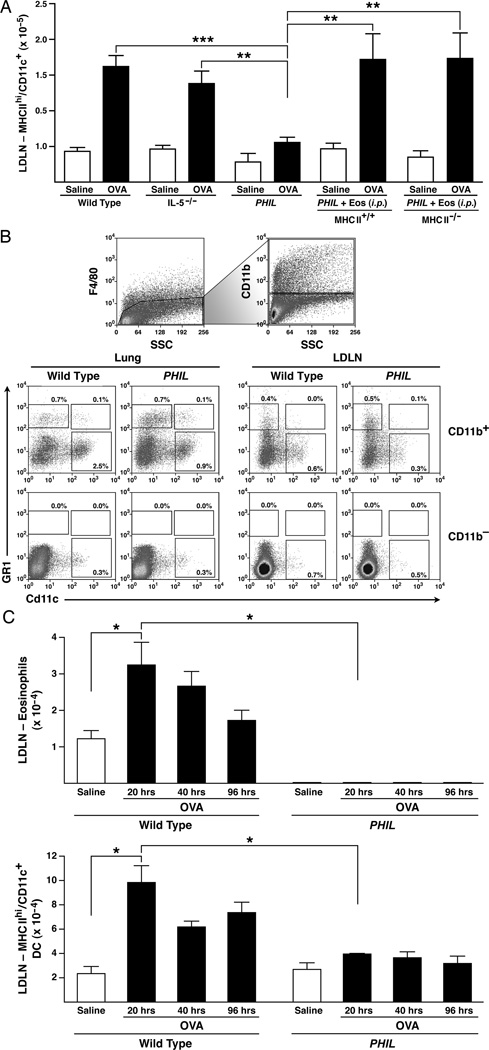
Reports have recently suggested that eosinophils have the potential to modulate allergen-dependent pulmonary immune responses. The studies presented expand these reports demonstrating in the mouse that eosinophils are required for the allergen-dependent Th2 pulmonary immune responses mediated by dendritic cells (DCs) and T lymphocytes. Specifically, the recruitment of peripheral eosinophils to the pulmonary lymphatic compartment(s) was required for the accumulation of myeloid DCs in draining lymph nodes and, in turn, Ag-specific T effector cell production. These effects on DCs and Ag-specific T cells did not require MHC class II expression on eosinophils, suggesting that these granulocytes have an accessory role as opposed to direct T cell stimulation. The data also showed that eosinophils uniquely suppress the DC-mediated production of Th17 and, to smaller degree, Th1 responses. The cumulative effect of these eosinophil-dependent immune mechanisms is to promote the Th2 polarization characteristic of the pulmonary microenvironment after allergen challenge.


Conflict of interest statement

The authors have no conflicting financial interests.

Figures

Figure 1
Figure 1. Peripheral eosinophils are required for T cell activation in LDLNs of OVA-treated mice
(A) Wild type, IL-5 knockout (IL-5−/−), and PHIL mice were subjected to OVA sensitization on day 0 and 14, acute OVA challenge on days 24–26 (OVA-treated), and assessed on day 28. Control animals were treated with saline alone. CD4+/TCR-β+ T cell numbers were determined by flow cytometry of total cells in the lungs of OVA-treated mice. (B) Mice treated as in (A) were injected with BrdU (4 hours prior to sacrifice) and assessed for total TCR-β+/BrdU+ T cells by flow cytometry (i.e., cell proliferation). (C) Eosinophil and CD4+ T cell numbers in mice were determined (day 28) in OVA-treated wild type, PHIL, and PHIL mice that were also transferred (i.p.) with 4 × 10⁷ eosinophils (Eos (i.p.)) one hour prior to the first OVA challenge). Representative FACS plots are shown of eosinophils (Siglec-F+/CCR3+) in the lung, LDLNs, spleen, and a non-pulmonary lymphoid compartment (mesenteric lymph node (MLN)). Numbers above boxes indicate percent of eosinophils out of the total live population. CD4+/TCR-β+ T cell numbers were assessed by flow cytometry of total cells in the LDLNs (D) and lungs (E) of OVA-treated mice following eosinophil adoptive transfer. All data are derived from at least three independent experiments each with cohort sizes of 2–6 mice (Mean ± SEM.). *p<0.05, **p<0.01, and ***p<0.001. (unpaired two-tailed student’s t-test).
Figure 2
Figure 2. MHC II expression on eosinophils is not necessary for T cell activation
OVA-treated wild type and PHIL mice as well as OVA-treated PHIL mice following adoptive transfer (i.p.) of either 4 × 10⁷ MHC II+/+ or MHC II−/− eosinophils (Eos (i.p.)) one hour prior to the first OVA challenge (day24) were assessed on day 28 of the protocol; control animals were challenged with saline alone. (A) Representative FACS plots of LDLN cells demonstrate the presence of eosinophils (Siglec-F+/CCR3+). Numbers above boxes indicate percent of eosinophils out of the total live population. (B) Effector T cell (CD4+/TCR-β+/CD44hi) numbers were determined in LDLNs from OVA-treated mice following eosinophil adoptive transfer. All data are derived from at least three independent experiments each with cohort sizes of 2–4 mice (Mean ± SEM.). *p<0.05; **p<0.01. (unpaired two-tailed student’s t-test).
Figure 3
Figure 3. Eosinophils are required for activation/accumulation of myeloid DCs
(A) OVA-treated wild type and PHIL mice as well as OVA-treated PHIL mice following adoptive transfer (i.p.) of either 4 × 10⁷ MHC II+/+ or MHC II−/− (Eos (i.p.)) one hour prior to the first OVA challenge (day24) were assessed on day 28 of the protocol; control animals were challenged with saline alone. OVA-sensitized wild type, IL5−/−, and PHIL mice challenged with either OVA or saline are shown for comparison. LDLNs of OVA-treated recipient PHIL mice were assessed for the presence of MHC IIhiCD11c+DCs by flow cytometry as calculated from total single cell suspensions of LDLNs. (B) Lungs and LDLN from OVA-treated wild type and PHIL mice that did not undergo eosinophil adoptive transfer were assessed for various DC populations. Total cell suspensions were gated on the F4/80-negative (non-macrophage) population and then gated into CD11b+ and CD11b groups. Percents out of total populations of these were determined for lymphocytic DCs (F4/80/CD11b+/Gr1+/CD11c+), plasmacytoid DCs (F4/80/CD11b/Gr1+/CD11c+) and monocytic inflammatory DCs (F4/80/CD11b+/Gr1+/Cd11c), and myeloid DCs (F4/80/CD11b+/Gr1/CD11c+) populations. (C) Kinetic assessment of eosinophils and activated DCs (MHC IIhiCD11c+) accumulation in the LDLNs of OVA-treated wild type vs. PHIL mice. (B). DC and eosinophil numbers were determined by collecting LDLN for flow cytometry analysis at 20, 40, and 96 hours after the first challenge of the three OVA challenges. All data are derived from at least three independent experiments each with cohort sizes of 2–5 mice (Mean ± SEM.). *p<0.05, **p<0.01, and ***p<0.001. (unpaired two-tailed student’s t-test).
Figure 4
Figure 4. OVA-pulsed myeloid DCs are sufficient for DC accumulation and T cell activation in the LDLNs of eosinophil-deficient mice
Bone marrow-derived and OVA-pulsed DCs (2–3 million cells) were transferred into the lungs of OVA-treated (OVA-sensitized/challenged) wild type or PHIL mice (i.t.) on day 24 of the acute OVA protocol and assessed on day 29. Mice receiving DC via i.t. transfer are indicated by (DC) and vehicle i.t. control is indicated by (Control). An additional cohort of OVA-treated PHIL mice also were adoptively transferred with 4 × 10⁷ eosinophils i.p. on the same day (i.e., day 24) as the DC transfer (DC + Eos). Non-sensitized but OVA-challenged PHIL recipients adoptively transferred (i.t.) with DCs (NS-PHIL) were included as controls to determine the necessity of antigen-dependent memory T cell activation. OVA sensitized/challenged wild type and PHIL mice subjected to the acute OVA protocol without cell transfer were performed as additional controls. (A) DCs (Gr1CD11c+) and (B) Effector (CD4+/TCRβ+/CD44hi) T cell populations in LDLNs were assessed in each group of mice by flow cytometry. All data are derived from at least four independent experiments each with cohort sizes of 2–4 mice (Mean ± SEM). *p<0.05 and **p<0.01 (unpaired two-tailed student’s t-test).
Figure 5
Figure 5. Transfer of OVA-pulsed myeloid DCs into PHIL leads to neutrophilic inflammation
Bone marrow-derived and OVA-pulsed DCs (2–3 million cells) were transferred into the lungs of OVA-sensitized/challenged wild type and PHIL mice by intratracheal instillation (+ DC (i.t.)) on the first day of OVA challenge (day 24) and assessed on day 29. An additional cohort of OVA-treated PHIL mice also were adoptively transferred with 4 × 10⁷ eosinophils i.p. on the same day (i.e., day 24) as the DC transfer (+ DC (i.t.) + Eos (i.p.)). In addition, bone-marrow derived OVA-pulsed myeloid DCs were transferred (i.t.) into a cohort of OVA-naïve recipient PHIL mice that were subsequently OVA-challenged (Non-sensitized/OVA-challenged (NS-PHIL)) to determine the necessity of antigen-dependent memory T cell activation. OVA sensitized/challenged wild type and PHIL mice subjected to the acute OVA protocol without cell transfer were performed as additional controls. (A) Representative images of lung sections assessing cellular inflammation (hematoxylin-eosin staining) and goblet cell metaplasia/airway epithelial cell mucin accumulation (PAS staining - dark purple cells). (B) Airway cellular infiltration was measured by calculating total bronchoalveolar fluid (BAL) cellularity and differentials of neutrophils, lymphocytes, and eosinophils. Mice receiving DC via (i.t.) transfer are indicated by (DC) and co-transfer of DCs (i.t.) and eosinophils (i.p.) is indicated by (DC + Eos) and vehicle transfer is indicated by (Control). Non-sensitized/OVA-challenged PHIL recipient mice + DC (i.t.) are indicated by (NS-PHIL). (C) BAL levels of IL-13 were measured from each group of mice. IL-17 and FN-γ levels were also performed these assessments and demonstrated that neither cytokine was detectable in the BALs of these mice. All data are derived from at least four independent experiments each with cohort sizes of 1–4 mice (Mean ± SEM). *p<0.05, **p<0.01, and ***p<0.001. (unpaired two-tailed student’s t-test).
Figure 6
Figure 6. Eosinophil-dependent suppression of Th17 immune responses following transfer of OVA-pulsed myeloid DCs
Bone marrow-derived and OVA-pulsed DCs (2–3 million cells) were transferred into the lungs of OVA-treated (OVA-sensitized/challenged) wild type or PHIL mice on day 24 of the acute OVA protocol described and assessed on day 29. Mice receiving DC via i.t. transfer are indicated by (DC) and transfer of vehicle alone is indicated by (Control). An additional cohort of OVA-treated PHIL mice also were adoptively transferred with 4 × 10⁷ eosinophils i.p. on the same day (i.e., day 24) as the DC transfer (DC + Eos). Non-sensitized but OVA-challenged PHIL recipients adoptively transferred (i.t.) with DCs are indicated by NS-PHIL. LDLN-derived cells from each cohort of mice were incubated for 72 hours in the presence of OVA and culture supernatant levels of IL-17, IFN-γ, and IL-13 were assessed by ELISA. All data are derived from at least four independent experiments each with cohort sizes of 2–4 mice (Mean ± SEM). *p<0.05 (unpaired two-tailed student’s t-test).

References

    1. Robinson DS. The role of the T cell in asthma. J Allergy Clin Immunol. 2010;126:1081–1091. quiz 1092-1083. - PubMed
    1. Bousquet J, Chanez P, Lacoste JY, Barneon G, Ghavanian N, Enander I, Venge P, Ahlstedt S, Simony-Lafontaine J, Godard P, Francois-Bernard M. Eosinophilic inflammation in asthma [see comments] New England Journal of Medicine. 1990;323:1033–1039. - PubMed
    1. MacKenzie JR, Mattes J, Dent LA, Foster PS. Eosinophils promote allergic disease of the lung by regulating CD4+ Th2 lymphocyte function. J Immunol. 2001;167:3146–3155. - PubMed
    1. Mattes J, Yang M, Mahalingam S, Kuehr J, Webb DC, Simson L, Hogan SP, Koskinen A, McKenzie AN, Dent LA, Rothenberg ME, Matthaei KI, Young IG, Foster PS. Intrinsic defect in T cell production of interleukin (IL)-13 in the absence of both IL-5 and eotaxin precludes the development of eosinophilia and airways hyperreactivity in experimental asthma. J Exp Med. 2002;195:1433–1444. - PMC - PubMed
    1. Jacobsen EA, Ochkur SI, Pero RS, Taranova AG, Protheroe CA, Colbert DC, Lee NA, Lee JJ. Allergic Pulmonary Inflammation in Mice is Dependent on Eosinophil-induced Recruitment of Effector T Cells. Journal of Experimental Medicine. 2008;205:699–710. - PMC - PubMed

Publication types