. 2011 Dec 1;187(11):5795-804.
doi: 10.4049/jimmunol.1102242. Epub 2011 Nov 2.

Lineage(-)Sca1+c-Kit(-)CD25+ cells are IL-33-responsive type 2 innate cells in the mouse bone marrow

Affiliations

Lineage(-)Sca1+c-Kit(-)CD25+ cells are IL-33-responsive type 2 innate cells in the mouse bone marrow

Adipong Brickshawana et al. J Immunol. .

Abstract

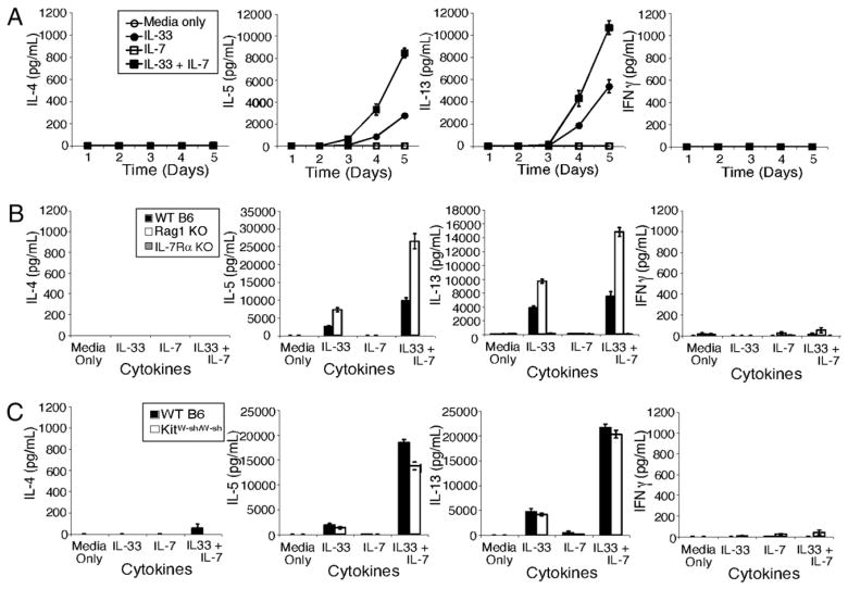
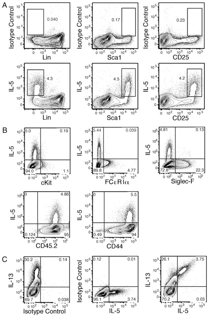
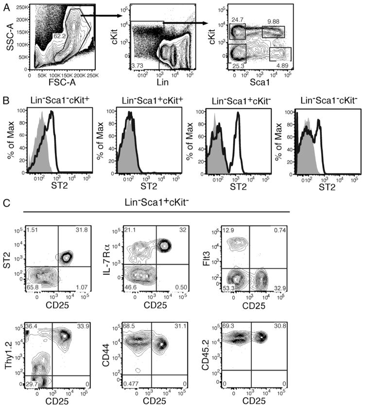
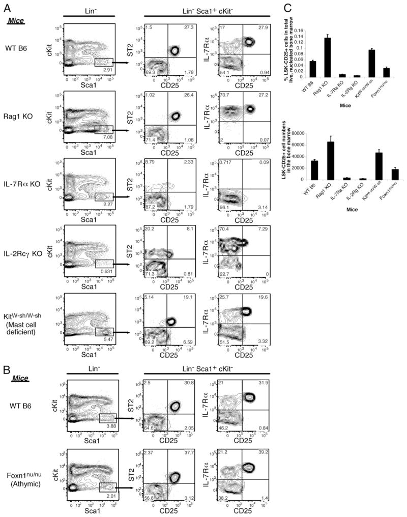
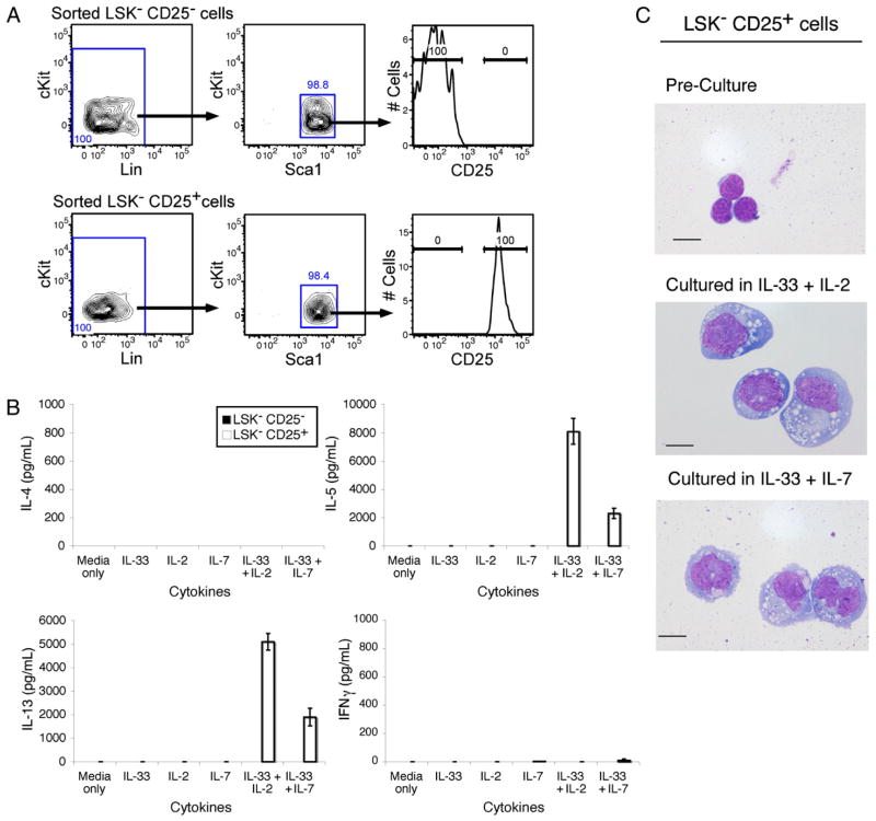
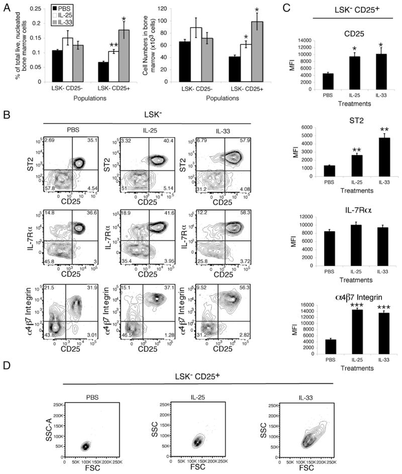
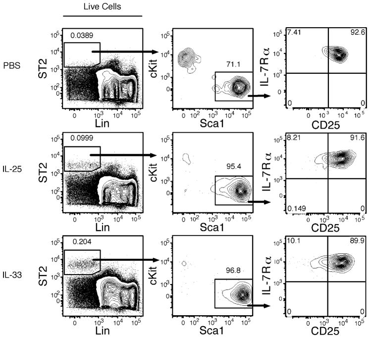
IL-33 promotes type 2 immune responses, both protective and pathogenic. Recently, targets of IL-33, including several newly discovered type 2 innate cells, have been characterized in the periphery. In this study, we report that bone marrow cells from wild-type C57BL/6 mice responded with IL-5 and IL-13 production when cultured with IL-33. IL-33 cultures of bone marrow cells from Rag1 KO and Kit(W-sh/W-sh) mice also responded similarly; hence, eliminating the possible contributions of T, B, and mast cells. Rather, intracellular staining revealed that the IL-5- and IL-13-positive cells display a marker profile consistent with the Lineage(-)Sca-1(+)c-Kit(-)CD25(+) (LSK(-)CD25(+)) cells, a bone marrow cell population of previously unknown function. Freshly isolated LSK(-)CD25(+) cells uniformly express ST2, the IL-33 receptor. In addition, culture of sorted LSK(-)CD25(+) cells showed that they indeed produce IL-5 and IL-13 when cultured with IL-33 plus IL-2 and IL-33 plus IL-7. Furthermore, i.p. injections of IL-33 or IL-25 into mice induced LSK(-)CD25(+) cells to expand, in both size and frequency, and to upregulate ST2 and α(4)β(7) integrin, a mucosal homing marker. Thus, we identify the enigmatic bone marrow LSK(-)CD25(+) cells as IL-33 responsive, both in vitro and in vivo, with attributes similar to other type 2 innate cells described in peripheral tissues.


Conflict of interest statement

Disclosures

The authors have no financial conflicts of interest.

Figures

FIGURE 1
FIGURE 1
Cytokine production from mouse bone marrow cells in response to IL-33 and IL-7. A, Bone marrow cells were harvested from C57BL/6 mice and cultured in media only or media supplemented with 10 ng/ml IL-33, 10 ng/ml IL-7, or 10 ng/ml IL-33 + 10 ng/ml IL-7. Culture supernatants were sampled daily, and IL-4, IL-5, IL-13, and IFN-γ levels were measured by ELISA. Error bars represent the SEM calculated from triplicate wells. B, Bone marrow cells from C57BL/6, Rag-1−/ −, and IL-7Rα −/ − mice were cultured as in A, and day 5 supernatants were analyzed by ELISA. Error bars represent the SEM calculated from 9 wells in three independent experiments. C, Bone marrow cells from C57BL/6 and KitW-sh/W-sh mice were cultured as in A, and day 5 supernatants were analyzed by ELISA. Error bars represent the SEM calculated from 6 wells in two independent experiments.
FIGURE 2
FIGURE 2
IL-5– and IL-13–producing cells in an in vitro culture of mouse bone marrow cells in media containing IL-33 + IL-7 are positive for progenitor and hematopoietic markers. Bone marrow cells from C57BL/6 mice were harvested as in Fig. 1 and were cultured in media supplemented with 10 ng/ml IL-33 + 10 ng/ml IL-7. BD GolgiPlug was added to culture media 12 h before intracellular cytokine staining and subsequent analysis by flow cytometry. Cells were stained for intracellular IL-5, or with corresponding isotype control Ab, and for surface expression of lineage markers (B220, CD3ε, CD4, CD8α, NK1.1, CD11b, CD11c, Gr-1, TER-119), Sca-1, and CD25 (A); or c-Kit, FcεRIα, SiglecF, CD45.2, or CD44 (B); or intracellular IL-13 (C). Gates in B and C were set using data from stains with corresponding isotype control Abs. All data shown are representative of at least two independent experiments.
FIGURE 3
FIGURE 3
ST2 expression on lineage low populations in the bone marrow. Bone marrow cells from 8-wk-old C57BL/6 mice were analyzed by flow cytometry. A, Gating strategy. Lineage low cells (low to no expression of B220, CD3ε, CD4, CD8α, DX5, CD11b, CD11c, Gr-1, and TER-119) were subdivided into four populations, based on their expression of Sca-1 and c-Kit. B, Expression of ST2 on the various lineage low subpopulations. Open histograms show anti-ST2 staining, and gray shaded histograms show isotype control Ab staining. Plots representative of stains from three different mice. C, ST2 and other cell surface markers expression on CD25 and CD25+LSK cells. Quadrants were drawn on the basis of isotype control Ab stains. All data shown are representative of stains from two to three mice.
FIGURE 4
FIGURE 4
LSKCD25+ cells in mutant and knockout mice. A, Bone marrow cells were harvested from 8- to 11-wk-old WT C57BL/6, Rag-1 KO, IL-7Rα KO, IL-2Rγ KO, and KitW-sh/W-sh or from 8- to 9-wk-old WT C57BL/6 and Foxn1nu/nu mice (B). Cells were stained as in Fig. 3 for LSKCD25+ cells and for their expression of ST2 and IL-7Rα. Plots representative of two to three mice per mouse type. C, The frequency and absolute cell number of LSKCD25+ cells in the bone marrow from two femurs and two tibias of each mouse. Error bars represent the SEM calculated from two to three mice per mouse type.
FIGURE 5
FIGURE 5
Sorted LSKCD25+ cells cultured in IL-33 + IL-2 and IL-33 + IL-7 produce IL-5 and IL-13 and lose their lymphoid morphology. Bone marrow cells from C57BL/6 mouse were lineage depleted using the Lineage Cell Depletion Kit (mouse) from Miltenyi Biotec. Lineage-depleted cells were stained and sorted for LSKCD25 and LSKCD25+ cells. A, Purity of sorted cells. B, Sorted cells were cultured in media only or media supplemented with 10 ng/ml IL-33, 10 ng/ml IL-2, 10 ng/ml IL-7, 10 ng/ml IL-33 plus 10 ng/ml IL-2, or 10 ng/ml IL-33 plus 10 ng/ml IL-7. Culture supernatants were harvested on day 5 of culture, and IL-4, IL-5, IL-13, and IFNγ productions were assayed by ELISA. Error bars represent the SEM calculated from 3 wells per condition. C, Wright–Giemsa stained LSKCD25+ cells preculture and after 5 d culture in media supplemented with IL-33 plus IL-2 or IL-33 plus IL-7. Photos were taken with ×1000 magnification. Scale bars, 10 μm. All data shown are representative of three independent experiments.
FIGURE 6
FIGURE 6
LSKCD25+ cells are responsive to IL-25 and IL-33 stimulation in vivo. PBS, 400 ng IL-25, or 400 ng IL-33 in PBS was injected i.p. into 8-wk-old WT C57BL/6 mice daily for 4 consecutive days. On day 5, nucleated bone marrow cells were harvested and stained for flow cytometry as in Fig. 3. A, Mean frequency and absolute cell number of LSKCD25 and LSKCD25+ cells per mouse in each treatment group. Error bars represent the SEM from four mice per treatment group (*p < 0.05, **p < 0.01 compared with PBS treatment by unpaired t test with Welch correction). B, ST2, IL-7Rα, and α4β7 integrin expression on LSKCD25 and LSKCD25+ cells across treatments. C, Mean fluorescence intensity (MFI) of CD25, ST2, IL-7Rα, and α4β7 integrin expression on LSKCD25+ cells across treatments. Error bars represent the SEM calculated from four mice per treatment group (*p < 0.05, **p < 0.01, ***p < 0.001 compared with PBS treatment by unpaired t test with Welch correction). D, Forward scatter and side scatter of LSKCD25+ cells across treatments. All data shown in B and D are representative of data from four mice per treatment group in two independent experiments.
FIGURE 7
FIGURE 7
ST2 high cells in the bone marrow of mice treated with IL-25 or IL-33 are LSKCD25+ cells. Total live bone marrow cells from Fig. 6 were analyzed for ST2 expression. Subgating on the ST2 high population shows that they are mostly LSK CD25+ cells. Plots are representative of data from four mice per treatment group in two independent experiments.

References

    1. Schmitz J, Owyang A, Oldham E, Song Y, Murphy E, McClanahan TK, Zurawski G, Moshrefi M, Qin J, Li X, et al. IL-33, an interleukin-1-like cytokine that signals via the IL-1 receptor-related protein ST2 and induces T helper type 2-associated cytokines. Immunity. 2005;23:479–490. - PubMed
    1. Liew FY, Pitman NI, McInnes IB. Disease-associated functions of IL-33: the new kid in the IL-1 family. Nat Rev Immunol. 2010;10:103–110. - PubMed
    1. Baekkevold ES, Roussigné M, Yamanaka T, Johansen FE, Jahnsen FL, Amalric F, Brandtzaeg P, Erard M, Haraldsen G, Girard JP. Molecular characterization of NF-HEV, a nuclear factor preferentially expressed in human high endothelial venules. Am J Pathol. 2003;163:69–79. - PMC - PubMed
    1. Carriere V, Roussel L, Ortega N, Lacorre DA, Americh L, Aguilar L, Bouche G, Girard JP. IL-33, the IL-1–like cytokine ligand for ST2 receptor, is a chromatin-associated nuclear factor in vivo. Proc Natl Acad Sci USA. 2007;104:282–287. - PMC - PubMed
    1. Moussion C, Ortega N, Girard JP. The IL-1-like cytokine IL-33 is constitutively expressed in the nucleus of endothelial cells and epithelial cells in vivo: a novel ‘alarmin’? PLoS One. 2008;3:e3331. - PMC - PubMed

Publication types

MeSH terms