. 2011 Dec 1;187(11):5703-11.
doi: 10.4049/jimmunol.1100558. Epub 2011 Nov 2.

A critical role for macrophages in promotion of urethane-induced lung carcinogenesis

Affiliations

A critical role for macrophages in promotion of urethane-induced lung carcinogenesis

Rinat Zaynagetdinov et al. J Immunol. .

Abstract

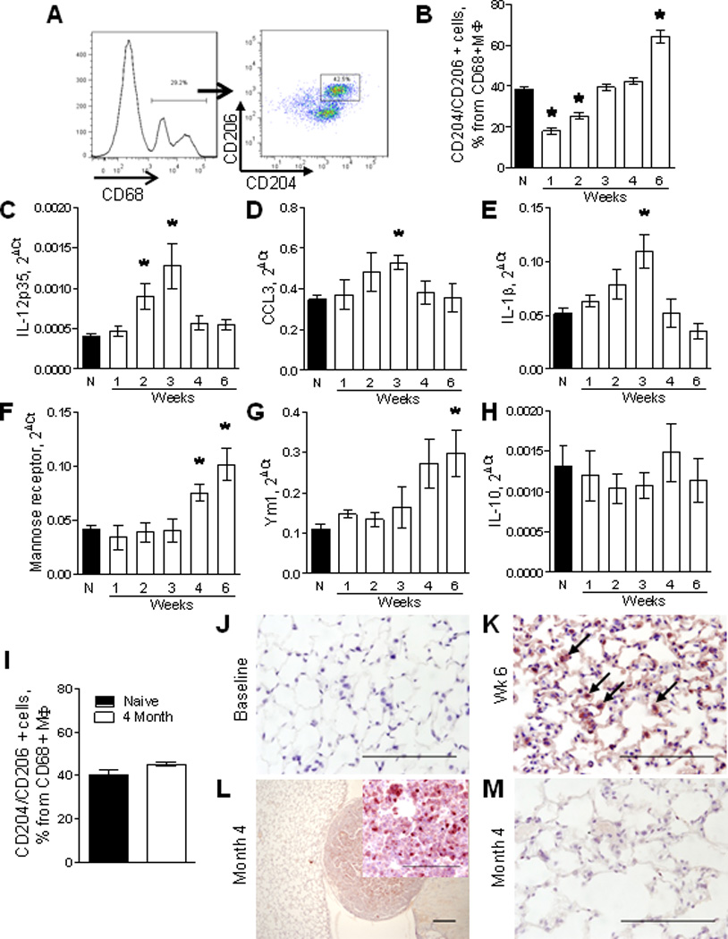
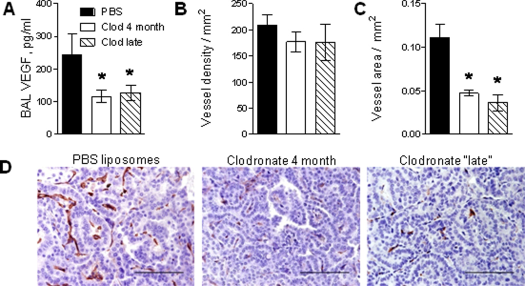
Macrophages have established roles in tumor growth and metastasis, but information about their role in lung tumor promotion is limited. To assess the role of macrophages in lung tumorigenesis, we developed a method of minimally invasive, long-term macrophage depletion by repetitive intratracheal instillation of liposomal clodronate. Compared with controls treated with repetitive doses of PBS-containing liposomes, long-term macrophage depletion resulted in a marked reduction in tumor number and size at 4 mo after a single i.p. injection of the carcinogen urethane. After urethane treatment, lung macrophages developed increased M1 macrophage marker expression during the first 2-3 wk, followed by increased M2 marker expression by week 6. Using a strategy to reduce alveolar macrophages during tumor initiation and early promotion stages (weeks 1-2) or during late promotion and progression stages (weeks 4-16), we found significantly fewer and smaller lung tumors in both groups compared with controls. Late-stage macrophage depletion reduced VEGF expression and impaired vascular growth in tumors. In contrast, early-stage depletion of alveolar macrophages impaired urethane-induced NF-κB activation in the lungs and reduced the development of premalignant atypical adenomatous hyperplasia lesions at 6 wk after urethane injection. Together, these studies elucidate an important role for macrophages in lung tumor promotion and indicate that these cells have distinct roles during different stages of lung carcinogenesis.


Figures

Figure 1
Figure 1
Depletion of alveolar macrophages attenuates urethane-induced lung tumorigenesis. A) Number of total cells in BAL from mice injected with urethane only (Ureth) or treated with weekly intratracheal (IT) injections of empty (PBS) liposomes or clodronate (Clod) liposomes for 4 month after urethane. B) Number of lung surface tumors and C) tumor diameter in mouse lungs. D) Histological assessment of the number of tumors per H&E stained lung section from tumor bearing mice treated with weekly IT injections of PBS liposomes or clodronate liposomes for 4 months. Data are presented as mean ± SEM of 6 mice for the urethane only group, 6 mice for the PBS group, and 10 mice for the clodronate group, *= p<0.05 compared to the other groups.
Figure 2
Figure 2
Characterization of macrophage phenotype in lungs following urethane treatment. A) Representative FACS plots demonstrating identification of M2 polarized macrophages in whole lungs of a naïve (untreated) mouse using anti-CD204 (scavenger receptor) and anti-CD206 (mannose receptor) antibodies. B) Analysis of the percentage of CD204 and CD206 double-positive cells within CD45+CD68+ macrophages in lungs of naïve mice (N) and mice at week 1–6 after urethane treatment. C–H) mRNA expression for M1 markers IL-12p35, CCL3, IL-1β and M2 markers mannose receptor, Ym1, IL-10 in total CD11b-positive cells isolated from lungs of naïve mice and 1–6 weeks after a single injection of urethane. n=4 mice per time point, *= p<0.05 compared to naïve mice. I) Percentage of CD204 and CD206 double-positive cells within lung CD45+CD68+ macrophages at 4 months after urethane injection. n=4 mice per group. J–M) Representative photomicrographs of lung sections stained with anti-arginase-1 antibodies at different time points after injection of urethane. Bars: 100µM.
Figure 3
Figure 3
Depletion of lung macrophages during “early” and “late” phases of lung tumorigenesis reduces tumor number and size. A) Schematic representation of “early” and “late” stage macrophage depletion experiment. B) The number of total BAL cells and C) alveolar macrophages in BAL of urethane-injected mice treated with clodronate or PBS liposomes. D) The number of lung surface tumors, E) surface tumor diameter, F) tumor number per lung section, and G) AAH lesions per H&E stained lung section. Data are presented as mean ± SEM of 12 mice for the PBS group (early and late PBS control groups are combined) and 11 mice for each clodronate group, *= p<0.05 compared to PBS control group.
Figure 4
Figure 4
Depletion of alveolar macrophages reduces tumor angiogenesis. A) VEGF concentration in BAL from mice treated with weekly IT clodronate beginning at 4 weeks post-urethane (late stage) or throughout the 4 months period compared to control mice treated with PBS liposomes. B) Density of blood vessels in tumors and C) blood vessel area in tumors. The blood vessel density (or area) was assessed as the number (or area) of CD34+ endothelial cells per square millimeter of tumor. C) Representative photomicrographs of lung sections from tumor bearing mice immunostained for CD34. Bars: 100µM. N=10–12 mice per group, *= p<0.05 compared to PBS liposome controls.
Figure 5
Figure 5
Depletion of macrophages with liposomal clodronate attenuates activity of NF-κB and alters inflammatory mediator expression in the lungs after urethane. A) Time course of NF-κB-dependent lung bioluminescence in NF-κB reporter mice (NGL) treated weekly with PBS or clodronate liposomes. n=6 for PBS liposome and 10 for clodronate groups, *=p<0.05 compared to PBS liposome group at the same time point. B) Representative images for NF-κB-dependent lung bioluminescence, C) NF-κB-dependent luciferase activity (Relative light units, RLU) in lung homogenates, and D) representative photomicrographs of lung sections immunostained for GFP (Bars: 100µM., arrows point to GFP+ cells) at day 14 after urethane injection in mice treated with PBS or clodronate liposomes, n=5 per group. E–I) Expression of mRNA for IL-12p35, iNOS, IL-6, TNFα, and IL-10 in lung tissue at day 11 after urethane injection in mice treated with PBS or clodronate liposomes (n=5 per group, *=p<0.05). Animals were given PBS or clodronate liposomes on days 0 and 7 after injection of urethane.
Figure 6
Figure 6
Depletion of alveolar macrophages during “early stage” lung carcinogenesis reduces formation of atypical adenomatous hyperplasia (AAH) lesions. A) Schematic representation of macrophage depletion experiment. B) Mean number of AAH lesions for each mouse counted per H&E stained lung section (3 per mouse) from mice treated with PBS liposomes or clodronate at day 0 and 7 (week 1,2) or week 4 and 5 and harvested at week 6 after injection of urethane (n=5 mice per group, *=p<0.05 compared to PBS liposome group). C) Total BAL cells and D) differential cell counts at week 6 from untreated mice (Naïve), mice treated with clodronate liposomes alone on day 0 and 7 and urethane-treated mice injected with clodronate or PBS liposomes on day 0 and 7, n=4–5 per group, *=p<0.05 compared to WT naive group.

References

    1. Jemal A, Siegel R, Xu J, Ward E. Cancer statistics, 2010. CA Cancer J. Clin. 2010;60:277–300. - PubMed
    1. Coussens LM, Werb Z. Inflammatory cells and cancer: think different! J. Exp. Med. 2001;193:F23–F26. - PMC - PubMed
    1. Hanahan D, Weinberg RA. The hallmarks of cancer. Cell. 2000;100:57–70. - PubMed
    1. Gordon S. Alternative activation of macrophages. Nat. Rev. Immunol. 2003;3:23–35. - PubMed
    1. Coussens LM, Werb Z. Inflammation and cancer. Nature. 2002;420:860–867. - PMC - PubMed

Publication types