. 2011 Nov 15;108(46):18702-7.
doi: 10.1073/pnas.1109348108. Epub 2011 Nov 2.

Nonautonomous regulation of Drosophila midgut stem cell proliferation by the insulin-signaling pathway

Affiliations

Nonautonomous regulation of Drosophila midgut stem cell proliferation by the insulin-signaling pathway

Na Hyun Choi et al. Proc Natl Acad Sci U S A. .

Abstract

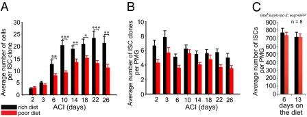
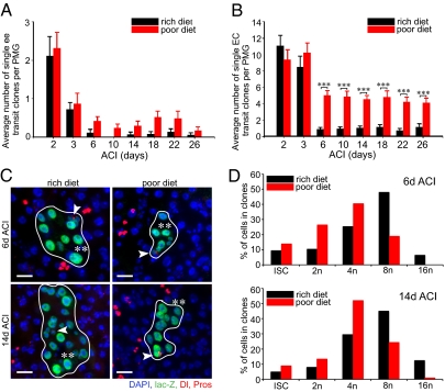
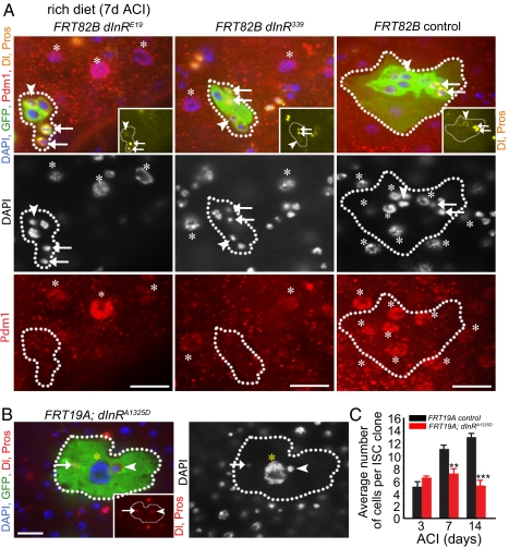
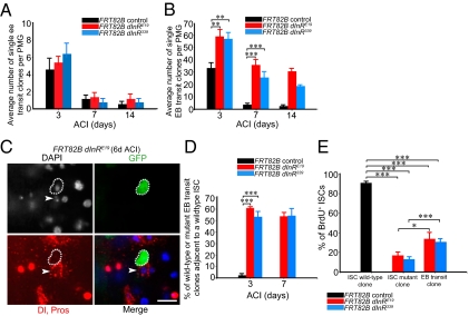
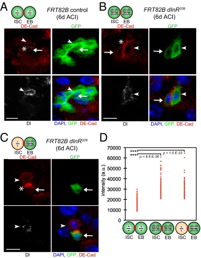
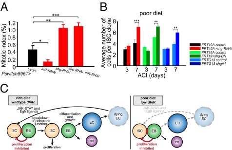
Drosophila adult midgut intestinal stem cells (ISCs) maintain tissue homeostasis by producing progeny that replace dying enterocytes and enteroendocrine cells. ISCs adjust their rates of proliferation in response to enterocyte turnover through a positive feedback loop initiated by secreted enterocyte-derived ligands. However, less is known about whether ISC proliferation is affected by growth of the progeny as they differentiate. Here we show that nutrient deprivation and reduced insulin signaling results in production of growth-delayed enterocytes and prolonged contact between ISCs and newly formed daughters. Premature disruption of cell contact between ISCs and their progeny leads to increased ISC proliferation and rescues proliferation defects in insulin receptor mutants and nutrient-deprived animals. These results suggest that ISCs can indirectly sense changes in nutrient and insulin levels through contact with their daughters and reveal a mechanism that could link physiological changes in tissue growth to stem cell proliferation.


Conflict of interest statement

The authors declare no conflict of interest.

Figures

Fig. 1.
Fig. 1.
Diet regulates adult posterior midgut homeostasis. (A) ISC clones from females fed a rich diet contain more cells than ISC clones from females fed a poor diet. (B) ISC clone number or (C) total posterior midgut stem cell number remains stable in females fed a rich or a poor diet. PMG, posterior midgut.
Fig. 2.
Fig. 2.
Diet regulates enterocyte growth. (A) Enteroendocrine transit clone turnover is unaffected in females fed a poor diet. (B) Enterocyte transit clone turnover decreases in females fed a poor diet. (C) Micrographs of ISC clones 6 d ACI and 14 d ACI from animals reared on a rich diet or a poor diet. Each clone contains a single ISC (arrowhead) and differentiating enterocytes. β-Galactosidase, green; Delta, red cytoplasmic vesicles; Prospero, red nuclear; and DAPI, blue nuclear. (Scale bar, 20 μm.) (D) Percentage of ISCs, 2n (non-ISCs), 4n, 8n, and 16n cells within ISC clones from females reared on a rich diet or a poor diet 6 and 14 d ACI. PMG, posterior midgut.
Fig. 3.
Fig. 3.
Insulin signaling regulates enterocyte differentiation. (A) dInRE19 mutant, dInR339 mutant, and control clones 7 d ACI from females reared on a rich diet. Clone, GFP green; Delta (yellow vesicular); Prospero (yellow nuclear); Pdm1 (red, nuclear) and DAPI (blue, nuclear). Asterisks mark mature enterocytes (Pdm1-positive cells). Dotted lines outline clone boundaries. (B) A clone overexpressing dInRA1325D in all cells contains a large enterocyte (yellow asterisks), an ISC (arrowhead), and an enteroendocrine cell (arrow). Clone, GFP green; Delta, red vesicular (Inset and merge); Prospero, red nuclear (Inset and merge); DAPI, blue nuclear. Arrowheads and arrows indicate, respectively, ISCs (Delta-positive) and enteroendocrine (Prospero-positive) cells in A and B. (Scale bar, 20 μm in A and B.) (C) Wild-type ISC clones contain more cells than ISC clones overexpressing an activated form of the insulin receptor (dInRA1325D).
Fig. 4.
Fig. 4.
Insulin signaling nonautonomously regulates intestinal stem cell proliferation. (A) Enteroendocrine dInR mutant transit clone and wild-type enteroendocrine transit clone turnover are similar. (B) Enteroblast dInR mutant transit clone turnover is decreased compared with wild-type enterocyte transit clone turnover. (C) A dInR mutant transit clone (green) in contact with a wild-type intestinal stem cell (arrowhead). Clone, GFP green; Delta, red vesicular; Prospero, red nuclear; DAPI, blue nuclear. (Scale bar, 20 μm.) (D) Percentage of mutant (FRT82B dInRE19, FRT82B dInR339) and wild-type (FRT82B) transit enteroblast clones adjacent to a wild-type ISC at 3 and 7 d ACI. (E) Percentage of BrdU incorporation in wild-type ISCs from wild-type clones and from wild-type ISCs adjacent to mutant transit clones 6 d ACI.
Fig. 5.
Fig. 5.
The insulin-signaling pathway regulates the stability of the adherens junction between ISCs and enteroblasts. (A) An FRT82B control ISC clone (green) 6 d ACI containing an ISC (arrowhead) and an enteroblast (arrow) adjacent (asterisk) to one another, as well as enterocytes. (B) A dInR339 mutant ISC clone (green) containing an ISC (arrowhead) and an enteroblast (arrow) in contact (asterisk). (C) A dInR339 mutant transit enteroblast clone (green, arrow) in contact (asterisk) with a wild-type intestinal stem cell (arrowhead). (AC) Clone, GFP green; Delta, white vesicular; DE–cadherin, red; DAPI, blue nuclear. (Scale bar, 10 μm.) (D) DE–cadherin levels measured at the junction of the ISC and the enteroblast are significantly higher in dInR339 mutant clones than in FRT82B clones.
Fig. 6.
Fig. 6.
Cell contact regulates ISC proliferation. (A) Expression of shg-RNAi in ISCs and enteroblasts suppresses proliferation defects caused by insulin receptor knockdown (dInR-RNAi). (B) shgIH MARCM clones, MARCM clones overexpressing shg-RNAi, or overexpressing shg-DN containing one stem cell from females reared on a poor diet grow larger than wild-type controls. (Scale bar, 20 μm.) (C) Model describing the nonautonomous role of nutrition and insulin signaling on ISC proliferation.

References

    1. Voog J, Jones DL. Stem cells and the niche: A dynamic duo. Cell Stem Cell. 2010;6:103–115. - PMC - PubMed
    1. Morrison SJ, Spradling AC. Stem cells and niches: Mechanisms that promote stem cell maintenance throughout life. Cell. 2008;132:598–611. - PMC - PubMed
    1. Ohlstein B, Spradling A. The adult Drosophila posterior midgut is maintained by pluripotent stem cells. Nature. 2006;439:470–474. - PubMed
    1. Micchelli CA, Perrimon N. Evidence that stem cells reside in the adult Drosophila midgut epithelium. Nature. 2006;439:475–479. - PubMed
    1. Cronin SJ, et al. Genome-wide RNAi screen identifies genes involved in intestinal pathogenic bacterial infection. Science. 2009;325:340–343. - PMC - PubMed

Publication types

LinkOut - more resources