. 2011 Nov 2;31(44):15861-9.
doi: 10.1523/JNEUROSCI.3272-11.2011.

Dynamic analysis of amyloid β-protein in behaving mice reveals opposing changes in ISF versus parenchymal Aβ during age-related plaque formation

Affiliations

Dynamic analysis of amyloid β-protein in behaving mice reveals opposing changes in ISF versus parenchymal Aβ during age-related plaque formation

Soyon Hong et al. J Neurosci. .

Abstract

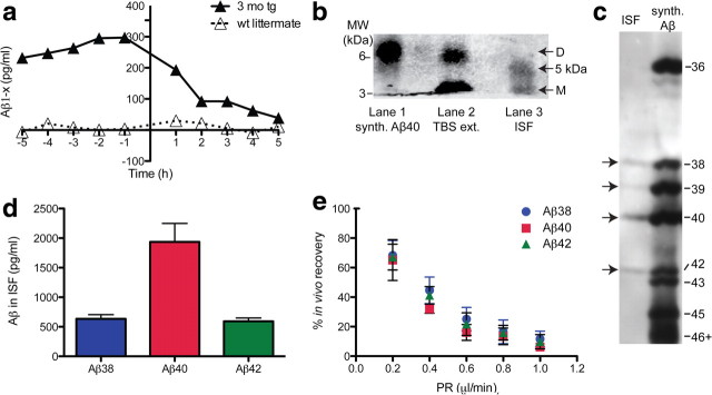
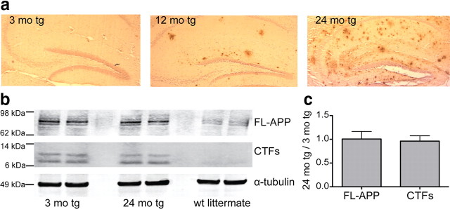
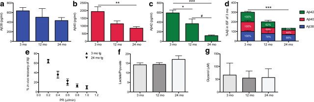
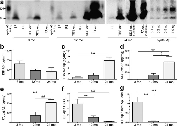
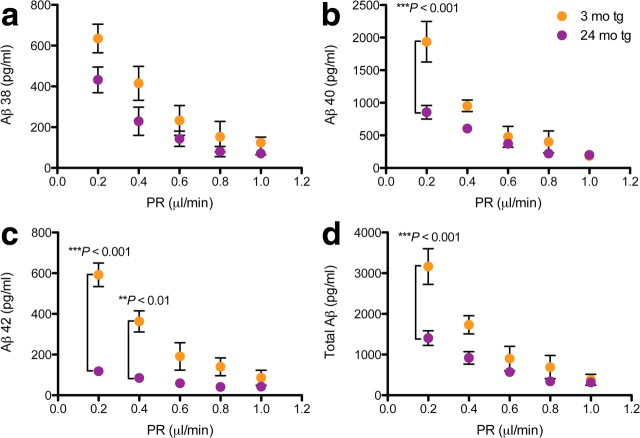
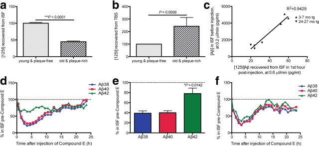
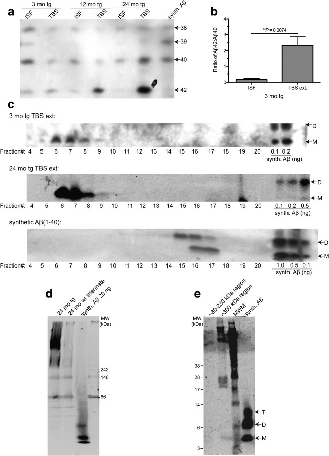
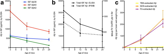
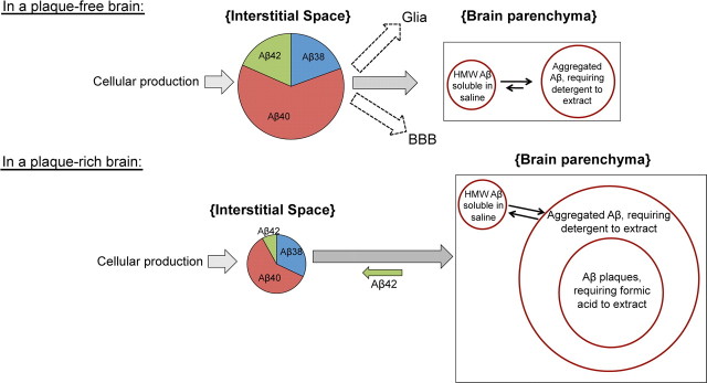
Growing evidence supports the hypothesis that soluble, diffusible forms of the amyloid β-peptide (Aβ) are pathogenically important in Alzheimer's disease (AD) and thus have both diagnostic and therapeutic salience. To learn more about the dynamics of soluble Aβ economy in vivo, we used microdialysis to sample the brain interstitial fluid (ISF), which contains the most soluble Aβ species in brain at steady state, in >40 wake, behaving APP transgenic mice before and during the process of Aβ plaque formation (age 3-28 months). Diffusible forms of Aβ, especially Aβ(42), declined significantly in ISF as mice underwent progressive parenchymal deposition of Aβ. Moreover, radiolabeled Aβ administered at physiological concentrations into ISF revealed a striking difference in the fate of soluble Aβ in plaque-rich (vs plaque-free) mice: it clears more rapidly from the ISF and becomes more associated with the TBS-extractable pool, suggesting that cerebral amyloid deposits can rapidly sequester soluble Aβ from the ISF. Likewise, acute γ-secretase inhibition in plaque-free mice showed a marked decline of Aβ(38), Aβ(40), and Aβ(42), whereas in plaque-rich mice, Aβ(42) declined significantly less. These results suggest that most of the Aβ(42) that populates the ISF in plaque-rich mice is derived not from new Aβ biosynthesis but rather from the large reservoir of less soluble Aβ(42) in brain parenchyma. Together, these and other findings herein illuminate the in vivo dynamics of soluble Aβ during the development of AD-type neuropathology and after γ-secretase inhibition and help explain the apparent paradox that CSF Aβ(42) levels fall as humans develop AD.


Figures

Figure 1.
Figure 1.
ISF Aβ obtained by microdialysis from behaving 3-month-old J20 hAPP tg mice. a, Rapid decline of ISF Aβ (t1/2, ∼2 h) upon acute γ-secretase inhibition in vivo in 3-month-old tg (vs wt littermate) mice. ISF sampled hourly at 1 μl/min; Compound E injected at time = 0 h. b, c, ISF collected at 0.2 μl/min were immunoprecipitated with AW8 Aβ antiserum and subjected to two types of SDS-PAGE. b, Conventional SDS-PAGE separates ISF Aβ into a ∼4 kDa [monomers (M)] and a ∼5 kDa Aβ-immunoreactive (lane 3) species. No dimers (D) were detected in ISF, but can be seen in the TBS-extract of a 24-month-old tg mouse (lane 2) or synthetic (synth.) Aβ (lane 1). WB was performed with 6E10 plus 4G8. c, Bicine/urea SDS-PAGE resolved ISF Aβ into three bands comigrating with synthetic Aβ1–38, Aβ1–40, and Aβ1–42, plus a fourth faint band corresponding to Aβ1–39. WB was performed with 6E10. d, Using 6E10 Aβ triplex ELISA, we quantified Aβx–38, Aβx–40, and Aβx–42 in ISF (mean ± SEM: 635 ± 70, 1937 ± 311, and 592 ± 58 pg/ml, respectively; n = 7 mice). e, Interpolated zero-flow method (mean ± SEM; n = 3–4 mice).
Figure 2.
Figure 2.
Amyloid plaques develop and mature with age in J20 APP tg mice without significant changes in full-length (FL) APP or in its proteolytic processing by β- or α-secretases. a, Hippocampal sections from fixed J20 APP tg brain were paraffin-embedded, then stained for Aβ using R1282 polyclonal antibody. Three-month-old tg sections were virtually plaque-free, whereas some plaques had formed by age 12 months. By 24 months, abundant diffuse and dense-core plaques populated the hippocampus. b, Representative blot of brain lysates of 3- and 24-month-old tg mice and wt littermate loaded onto denaturing SDS-PAGE, then blotted for full-length APP and its C-terminal fragments (CTFs) (WB was performed with polyclonal C7) or to α-tubulin (WB was performed with polyclonal tubulin-α). c, Summary ratios of immunoreactive signals at 24- versus 3-month-old tg mice, for full-length APP and C-terminal fragments normalized to the α-tubulin signal. n = 3 mice per group; signal quantification by Licor Odyssey.
Figure 3.
Figure 3.
Levels of soluble ISF Aβ <35 kDa in the brain fall with age. a–d, ISF was sampled from the hippocampi of 3- (preplaque), 12- (early plaque deposition), and 24-month old (abundant, mature plaques) J20 tg mice. Using Aβ triplex ELISAs, we found that Aβx–38 (a), Aβx–40 (b), and Aβx–42 (c) all decreased with age (means ± SEM; n = 7, 4, 7 mice at 3, 12, and 24 months, respectively). One-way ANOVA, followed by Bonferroni test: *p < 0.05, **p < 0.01, and ***p < 0.001 versus 3 months values; #p < 0.05 versus 12 months values. d, Proportional levels of Aβ38, Aβ40, and Aβ42, where each peptide was normalized to its level at 3 months. Aβ42 declined the most (80% by 24 months vs 3 months). ***p < 0.0001 by two-way ANOVA with age as a variant. e, At 0.2 μl/min, the perfusion rate used to collect all ISF samples in Figures 3 and 4, we obtained comparable percentage recoveries of microdialyzable Aβ in the two extreme ages (∼63 ± 3% in 3-month-old tg mice and ∼66 ± 2% in 24-month-old tg mice, means ± SEM; n = 3–4 mice). f, g, Ratios of lactate to pyruvate (f) and glycerol (g) levels in the ISF microdialysates were not altered with age (means ± SEM; n = 3–8 mice).
Figure 4.
Figure 4.
Aβ in all pools of brain parenchyma accrue with age while those that remain diffusible in the ISF declines. a, Representative IP/WBs of Aβ species from brains of the same mice right after microdialysis, in four pools: ISF, TBS extracted (ext), SDS extracted, and FA extracted. All pools (except FA extracted, which was lyophilized and straight-loaded onto the gel) were immunoprecipitated with AW8 and blotted with 6E10 and 4G8. Synthetic (synth.) Aβ run alongside for quantification. Perfusion buffer (PB) and TBS were immunoprecipitated as negative controls. b–e, Quantification of IP/WBs from 21 mice shows ∼50% decrease in absolute values of ISF Aβ between 3 and 12 months (not significant by one-way ANOVA followed by Bonferroni test) (b), with a sharp rise in TBS- (c), SDS- (d), and FA-(e) extracted Aβ (picograms per milligrams wet brain tissue). f, g, Ratios of ISF to TBS-soluble Aβ (f) or to total parenchymal Aβ (g) calculated for each mouse and shown as mean ratio ± SEM; n = 7 mice per group. Aβ quantified by Licor Odyssey imaging and analyzed by one-way ANOVA and Bonferroni test: **p < 0.01 and ***p < 0.001 versus 3 months; #p < 0.05 and ##p < 0.01 versus 12 months. D, Dimers; M, monomers.
Figure 5.
Figure 5.
Microdialysis at slower perfusion rates reveals age-dependent changes in ISF Aβ. ISF were collected from hippocampi of 3- (plaque-free) and 24- (plaque-rich) month-old tg mice while varying the PR. Samples were then quantified using 6E10 Aβ triplex ELISA. a–c, At PR 1 μl/min, no age-dependent changes in any of the three peptides were detectable; however, at slow PRs (especially 0.2 μl/min), significant differences were noted, particularly in Aβ40 and Aβ42. d, Sum of the three Aβ peptides measured (total Aβ). Values are means ± SEM, n = 3–7 mice; p values by two-way ANOVA and Bonferroni test.
Figure 6.
Figure 6.
Dynamic shift in the in vivo economy of Aβ once plaques develop. a–c, Soluble synthetic [125I]Aβ1–40 at a physiological concentration (1 nm) was injected intrahippocampally via a small cannula on the microdialysis probe in awake, behaving mice at ages 3–7 (plaque-free) or 24–27 (plaque-rich) months, and radioactivity was recovered in the ISF by microdialysis (a) and from the TBS extracts of the brains (b). Means normalized to amounts in the plaque-free mice (100%) ± SEM; n = 3–4 mice per group; p values are one-tailed t tests versus 3–7 months. c, Amount of [125I]Aβ recovered in the first hour of injection plotted versus the endogenous Aβ levels sampled from same mice before injection. d–f, ISF Aβ42 is minimally affected by Compound E in plaque-rich mice. d, Representative graph of hourly ISF Aβ levels after Compound E injection (0 h) into 24-month-old plaque-rich mice while collecting their brain ISF at 0.6 μl/min. Levels were normalized to levels of each Aβ species before injection. e, Quantification of the individual ISF Aβ peptides in 24-month-old plaque-rich mice for the first 5 h postinjection (means ± SEM, n = 3 mice; p value is by one-way ANOVA). f, Hourly ISF Aβ levels after Compound E injection (0 h) in a 3-month-old plaque-free tg mouse.
Figure 7.
Figure 7.
Saline brain extracts of tg mouse exists principally in assemblies >500 kDa in native form. a, Bicine/urea SDS-PAGE shows the principal Aβ peptides present and their relative expression levels in ISF and TBS extracts of 3-, 12-, and 24-month-old tg mice. IP was performed with AW8; WB was performed with 6E10. b, Quantification (by ImageJ software) of Aβ42/Aβ40 ratios in plaque-free 3-month-old tg mice (p value by one-tailed t test; n = 3 each). c, Non-denaturing SEC of TBS extracts (ext) of 3- and 24-month-old tg mice performed on a Superdex 200 SEC column followed by SDS-PAGE of each SEC fraction. Synthetic (synth.) Aβ40 run on the same SEC column for comparison. WB was performed with 3D6. d, TBS extracts from a 24-month-old tg mouse and its wt littermate subjected to clear native PAGE and blotted for Aβ. WB was performed with 2G3 plus 21F12. e, Excision of the >300 kDa region and subsequent electrophoresis by denaturing SDS-PAGE showed that this high MW material is disassembled into low MW SDS-stable Aβ species. WB was performed with 3D6. T, Trimers; D, dimers; M, monomers; MWM, MW marker.
Figure 8.
Figure 8.
Summary of the temporal changes in the four Aβ brain pools. a, Decreases with age in all three in vivo ISF Aβ peptides measured by 6E10 Aβ triplex sandwich ELISA (for details, see Fig. 3). b, The fold-decrease measured by ELISA (significant between ages 3 and 24 months by two-way ANOVA; see Fig. 2) is comparable to the fold-decrease measured by IP/WB (though the latter was not significant by one-way ANOVA; for details, see Fig. 4). The total amount measured by IP/WB analysis method was only ∼30% of the total amount measured by ELISA. c, IP/WB analysis of Aβ in brain tissue. By ∼24 months, there was a steep increase in insoluble (SDS- and FA-extracted) Aβ. The TBS-extracted Aβ from the same mice did not rise until 24 months and then only very slightly. Values are means ± SEM from Figures 3 and 4.
Figure 9.
Figure 9.
A hypothetical model of Aβ in vivo dynamics before versus after plaque formation based on data herein. Several factors contribute to steady-state Aβ levels in brain ISF. There is a constant supply of Aβ in the ISF pool generated by new APP processing (Aβ40 ≫ Aβ38 = Aβ42 > Aβ39), and this generation appears to change little with age. ISF Aβ can be proteolytically degraded, cleared locally by glia and/or transported across the blood brain barrier (BBB). Soluble Aβ starts aggregating at an early age in brain parenchyma, as evidenced by the >500 kDa TBS-extractable pool as well as an SDS-extractable pool in 3-month-old plaque-free mice. The most abundant pools in a plaque-free brain are the ISF pool and the SDS-extractable pool. In a plaque-rich brain, however, equilibrium between pools is greatly altered by the overwhelming amount of aggregated Aβ, which act as a sink, thereby diminishing the steady-state ISF pool. Plaques may also act as a contributor to the ISF pool, where Aβ42, the most abundant peptide in plaques (and also the most decreased in the ISF) diffuses back into the ISF. HMW, High MW (i.e., >500 kDa).

References

    1. Bech RA, Waldemar G, Gjerris F, Klinken L, Juhler M. Shunting effects in patients with idiopathic normal pressure hydrocephalus; correlation with cerebral and leptomeningeal biopsy findings. Acta Neurochir (Wien) 1999;141:633–639. - PubMed
    1. Brody DL, Magnoni S, Schwetye KE, Spinner ML, Esparza TJ, Stocchetti N, Zipfel GJ, Holtzman DM. Amyloid-beta dynamics correlate with neurological status in the injured human brain. Science. 2008;321:1221–1224. - PMC - PubMed
    1. Cirrito JR, May PC, O'Dell MA, Taylor JW, Parsadanian M, Cramer JW, Audia JE, Nissen JS, Bales KR, Paul SM, DeMattos RB, Holtzman DM. In vivo assessment of brain interstitial fluid with microdialysis reveals plaque-associated changes in amyloid-beta metabolism and half-life. J Neurosci. 2003;23:8844–8853. - PMC - PubMed
    1. Cirrito JR, Kang JE, Lee J, Stewart FR, Verges DK, Silverio LM, Bu G, Mennerick S, Holtzman DM. Endocytosis is required for synaptic activity-dependent release of amyloid-beta in vivo. Neuron. 2008;58:42–51. - PMC - PubMed
    1. Del Bigio MR, Cardoso ER, Halliday WC. Neuropathological changes in chronic adult hydrocephalus: cortical biopsies and autopsy findings. Can J Neurol Sci. 1997;24:121–126. - PubMed

Publication types

MeSH terms