. 2012 Aug;16(8):1840-55.
doi: 10.1111/j.1582-4934.2011.01479.x.

Involvement of COX-2/PGE2 signalling in hypoxia-induced angiogenic response in endothelial cells

Affiliations

Involvement of COX-2/PGE2 signalling in hypoxia-induced angiogenic response in endothelial cells

Lixing Zhao et al. J Cell Mol Med. 2012 Aug.

Abstract

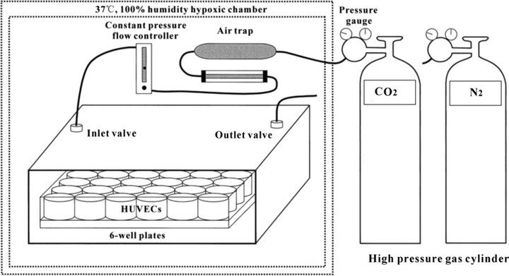
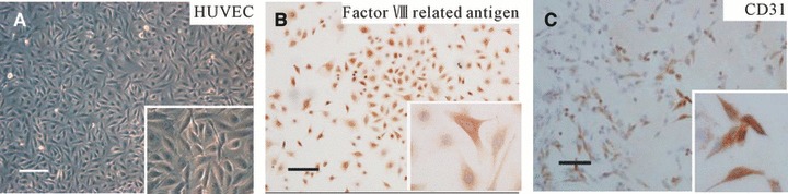
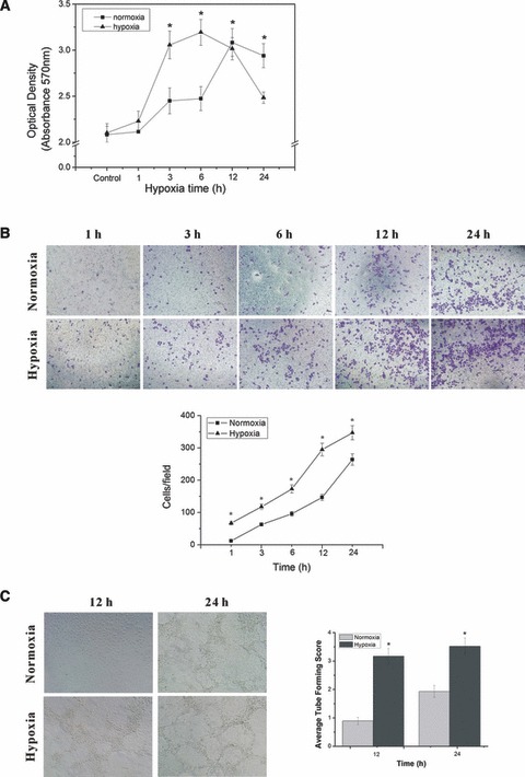
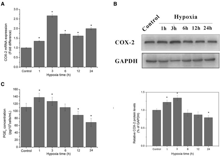
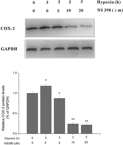
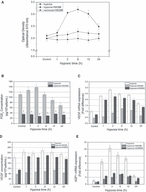
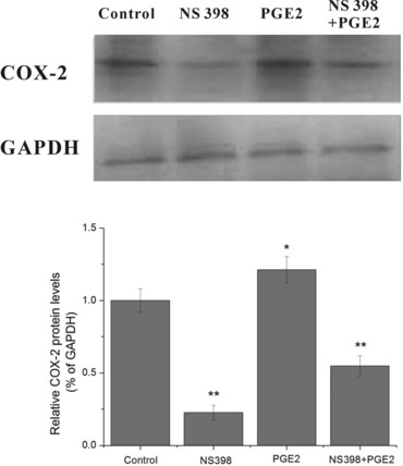
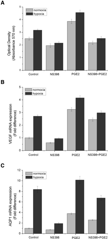
To evaluate the impact of hypoxia on the angiogenic capability of endothelial cells (ECs), and further investigate whether the cyclooxygenase-2 (COX-2)/prostaglandin E(2) (PGE(2)) signalling is involved in the angiogenic response of ECs to hypoxia. We explored the impact of various periods (1, 3, 6, 12, 24 hrs) of hypoxia (2% O(2)) on human umbilical vein endothelial cells (HUVECs) in vitro. We observed cell viability, migration, tube formation, analysed COX-2, vascular endothelial growth factor (VEGF), AQP1 mRNA transcription, protein expression and measured PGE(2), VEGF protein concentration in cell supernatants. Then we treated HUVECs with COX-2 selective inhibitor NS398, EP1/2 combined antagonist AH6809 and exogenous PGE(2) to investigate the role of COX-2/PGE(2) signalling in the angiogenic response of ECs to hypoxia. The results demonstrated that short-term hypoxic treatment enhanced HUVECs proliferation, migration, tube formation, significantly up-regulated COX-2, VEGF, AQP1 mRNA level, protein expression and promoted PGE(2) , VEGF release. The pharmacological inhibition study revealed that exposure of HUVEC to NS398 and AH6809 under hypoxia impaired the biological responses of ECs to hypoxia. Exogenous PGE(2) augments the effects of hypoxia on HUVECs, and partially reversed the inhibitory effects of NS398 on HUVECs proliferation and angiogenic capability. Short-term hypoxic treatment enhanced angiogenic capability of ECs, and COX-2/PGE(2) signalling may play a critical role in the biological response of ECs to hypoxia.


Figures

Fig 1
Fig 1
Schematic diagram of the Binder, a three gas modular hypoxic incubator used to simulate hypoxic conditions in vivo. Ambient oxygen concentrations of 2% were maintained using this device with CO2/O2 monitoring and CO2/N2 gas sources. A pure N2 entrance is set for the nitrogen replacement of air in incubator to control the oxygen concentration and achieve the purpose of hypoxia.
Fig 2
Fig 2
Characterization of HUVECs by morphological observation and immunocytochemical staining. (A) The cells exhibited typical cobblestone morphology on reaching confluence, insert with higher magnification of ×200. (B and C) Immunocytochemical stained with factor VIII-related antigen and CD31 antibody, respectively, the immunoreactive positive cells were stained brownish-yellow, insert with higher magnification of ×400 confirming brownish-yellow stained cytoplasm. Scale bars: 100 μm.
Fig 3
Fig 3
HUVECs proliferation, migration and tube formation under hypoxic exposure. (A) HUVECs proliferation and viability following hypoxic exposure by MTT assay. *P < 0.05 versus control group. (B) HUVECs were assayed for migration under normoxia or hypoxia for indicated periods of time. The cells migrate across the membrane were counted under a light microscope with an eyepiece grid to visualize set fields. At least four fields in duplicate wells were counted for each group to generate the bar chart. (C) The tube formation of HUVECs after 12 and 24 hrs of normoxia or hypoxia was viewed by phase-contrast microscopy at ×100 magnification, tube formation of each group was scored on a scale of 0–5 based on quality and number of the tubes, score results from four random fields in duplicate wells were averaged to generate the bar chart. *P < 0.05 versus normoxia group.
Fig 4
Fig 4
HUVECs proliferation, VEGF, AQP1 mRNA level, protein expression and VEGF accumulation in supernatant under hypoxic exposure. (A) The mRNA levels of VEGF at different hypoxic time points. (B) The protein expression of VEGF at different hypoxic time points by Western blotting analysis, the blots and the bar chart below are in one label. (C) Quantification of VEGF by ELISA assay in supernatant of HUVECs after various periods of hypoxic treatment. (D) The mRNA levels of AQP1 at different hypoxic time points. (E) The protein expression of AQP1 at different hypoxic time points by Western blotting analysis, the blots and the bar chart below are in one label. *P < 0.05 versus control group.
Fig 5
Fig 5
COX-2 mRNA level, protein expression and PGE2 accumulation in supernatant under hypoxic exposure. (A) The mRNA levels of COX-2 at different hypoxic time points. 2−ΔΔCt values were obtained by real-time RT-PCR analysis using GAPDH transcripts for the normalization. (B) The protein expression of COX-2 at different hypoxic time points by Western blotting analysis, the blots and the bar chart below are in one label. (C) Quantification of PGE2 by ELISA assay in supernatant of HUVECs after various periods of hypoxic treatment, the data are expressed in concentration of PGE2. *P < 0.05 versus control group.
Fig 6
Fig 6
Effect of the selective COX-2 inhibitor NS398 on COX-2 activity by concentration gradient test. HUVECs were exposed to hypoxia and supplemented with NS398 at indicated concentrations of 0, 5, 10 and 20 μM. Cells without hypoxia or NS398 treatment served as control. The COX-2 activity was determined by Western blot analysis. *P < 0.05, **P < 0.01 versus control.
Fig 7
Fig 7
Selectively inhibition of COX-2 activity by NS398 and blockade of EP1/2 by AH6809 impair the biological response of HUVECs to hypoxia. (A) HUVECs proliferation and viability following normoxic, hypoxic or hypoxic + NS398 treatment by MTT assay. (B) Quantification of PGE2 release by ELISA assay in supernatant of HUVECs after various periods of hypoxic treatment with or without 10 μM NS398. (C) VEGF mRNA level assessed by real-time RT-PCR experiment, HUVECs were exposed to hypoxia in the presence or absence of NS398 (10 μM) for indicated periods of time. (D) VEGF concentration in supernatant of HUVECs measured by ELISA assay. (E) AQP1 mRNA level assessed by real-time RT-PCR experiment. *P < 0.05 versus control.
Fig 8
Fig 8
Effect of NS398, exogenous PGE2 and combinational treatment of NS398 and exogenous PGE2 on COX-2 protein expression of HUVECs under normoxia. HUVECs were treated with 10 μM of NS398 and/or 10 μM of exogenous PGE2 under normoxia for 3 hrs. Cells without NS398 or exogenous PGE2 treatment served as control. The COX-2 protein expression was determined by Western blot analysis. *P < 0.05 and **P < 0.01 versus control.
Fig 9
Fig 9
Effect of NS398, exogenous PGE2 and combinational treatment of NS398 and exogenous PGE2 on cell viability and VEGF, AQP1 mRNA transcription of HUVECs under normoxia or hypoxia. HUVECs were treated with 10 μM of NS398 and/or 10 μM of exogenous PGE2 under normoxia or hypoxia for 3 hrs. Cells without NS398 or exogenous PGE2 treatment served as control. (A) HUVECs proliferation and viability was measured by MTT assay. (B) VEGF mRNA level assessed by real-time RT-PCR experiment. (C) AQP1 mRNA level assessed by real-time RT-PCR experiment. *P < 0.05 and **P < 0.01 versus control.

References

    1. Folkman J, Shing Y. Angiogenesis. J Biol Chem. 1992;267:10931–4. - PubMed
    1. Bernas GC. Angiotherapeutics from natural products: from bench to clinics? Clin Hemorheol Microcirc. 2003;29:199–203. - PubMed
    1. Ku SJ, Chang YI, Chae CH, et al. Static tensional forces increase osteogenic gene expression in three-dimensional periodontal ligament cell culture. BMB Rep. 2009;31:427–32. - PubMed
    1. Grellier M, Ferreira-Tojais N, Bourget C. Role of vascular endothelial growth factor in the communication between human osteoprogenitors and endothelial cells. J Cell Biochem. 2009;106:390–8. - PubMed
    1. Jain RK. Molecular regulation of vessel maturation. Nat Med. 2003;9:685–93. - PubMed

Publication types

MeSH terms