Case Reports
. 2012 Mar 15;314(1-2):130-7.
doi: 10.1016/j.jns.2011.10.006. Epub 2011 Nov 1.

Hereditary diffuse leukoencephalopathy with axonal spheroids (HDLS): a misdiagnosed disease entity

Affiliations
Case Reports

Hereditary diffuse leukoencephalopathy with axonal spheroids (HDLS): a misdiagnosed disease entity

Christina Sundal et al. J Neurol Sci. .

Abstract

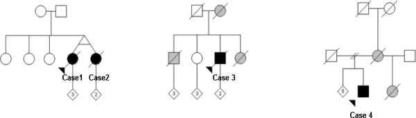
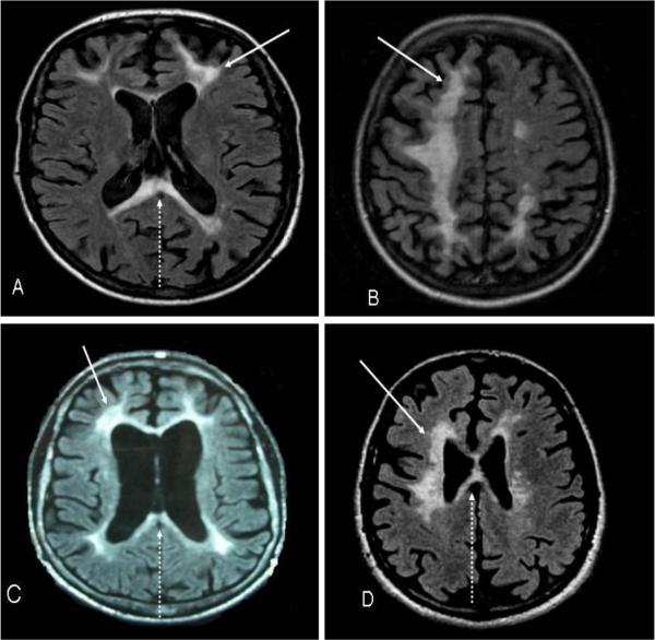
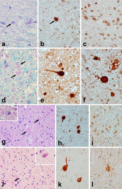
Hereditary diffuse leukoencephalopathy with spheroids (HDLS) was originally described in a large Swedish pedigree. Since then, 22 reports describing a total of 13 kindreds and 11 sporadic cases have been published. Inheritance is autosomal dominant, albeit the gene is unknown. Here we report on the clinical findings, genealogical data, brain MRI data, and autopsy/biopsy findings of four probands from three independently ascertained novel families from Norway, Germany and US. We identified a 39-year-old female and her twin sister, a 52-year-old male and a 47-year-old male with progressive neurological illness characterized by personality changes, cognitive decline and motor impairments, such as gait problems, bradykinesia, tremor and rigidity. Brain MRI showed white matter abnormalities with frontal prominence. Brain biopsy/autopsies were consistent with HDLS. HDLS is an under-recognized disease and in reporting these cases, we aim to increase the awareness of the disorder. Due to varied and wide phenotypic presentations, which may imitate several neurodegenerative diseases, HDLS can be difficult to diagnose. Definitive diagnosis can be established only by direct brain tissue examination. Familiarity with the clinical presentation and typical neuroimaging findings may be helpful in narrowing the diagnosis.


Figures

Fig. 1
Fig. 1. Pedigrees
Circles represent females and squares represent males. Black symbols indicate pathologically confirmed HDLS. Grey symbols indicate possible diagnosis. Arrow indicates the proband. Slash indicates deceased. Numbers inside the diagonal symbol indicates children. Cases 1 and 2: Norway; Case 3: Germany; Case 4: US (Polish and German descent).
Fig. 2
Fig. 2. Magnetic resonance images (axial sections, T2-weighted) from the 4 patients
(A)Case 1 (MRI performed 1.2 years after start of symptoms); localized white matter lesions (arrow) in both frontal and parietal hemispheres involving the corpus callosum (arrow dashed). (B)Case 2 (MRI performed 1.9 years after start of symptoms); confluent white matter lesions in both frontal and parietal hemispheres with cortical atrophy in the affected areas. (C)Case 3 (MRI performed 3.5 years after start of symptoms); localized periventricular lesions (arrow) with corresponding frontoparietal atrophy and involvement of the corpus callosum (arrow dashed). (D)Case 4 (MRI performed 2.5 years after start of symptoms); bilateral frontoparietal white matter changes (arrow) extending into the corpus callosum (arrow dashed).
Fig. 3
Fig. 3. Neuropathology
Case 1: (a) Luxol fast blue stain for myelin shows myelin loss and tissue vacuolation with axonal spheroids [arrows in (a) and (b)], that are immunopositive for amyloid precursor protein (b). Affected white matter has many lipid-laden macrophages immunohistochemistry-positive for HLA-DR (c). Case 2: (d) Luxol fast blue stain for myelin shows myelin loss and tissue vacuolation with axonal spheroids [arrows in (d)] that are immunopositive for amyloid precursor protein (e) and for phosphorylated neurofilament (f). Astrocytes in affected white matter are hypertrophic and bizarre appearing with immunohistochemistry for alpha-B-crystallin (l).Case 3: (g) Hematoxylin & eosin stain shows myelin pallor with pigment-containing macrophages (inset) and axonal spheroids [arrows] that are immunopositive for amyloid precursor protein (h). Astrocytes in affected white matter are hypertrophic and bizarre appearing with immunohistochemistry for alpha-B-crystallin (i). The cortex overlying areas with white matter pathology frequently has ballooned / swollen neurons best appreciated with immunohistochemistry for alpha-B-crystallin (k). Case 4: (j) Hematoxylin & eosin stain of the brain biopsy shows myelin pallor with pigment-containing macrophages (inset) and axonal spheroids [arrow]. [All images are originally ×400]

References

    1. Axelsson R, Roytta M, Sourander P, Akesson HO, Andersen O. Hereditary diffuse leucoencephalopathy with spheroids. Acta Psychiatr Scand Suppl. 1984;314:1–65. - PubMed
    1. Wider C, Van Gerpen JA, DeArmond S, Shuster EA, Dickson DW, Wszolek ZK. Leukoencephalopathy with spheroids (HDLS) and pigmentary leukodystrophy (POLD): a single entity? Neurology. 2009;72:1953–9. - PMC - PubMed
    1. Van Gerpen JA, Wider C, Broderick DF, Dickson DW, Brown LA, Wszolek ZK. Insights into the dynamics of hereditary diffuse leukoencephalopathy with axonal spheroids. Neurology. 2008;71:925–9. - PMC - PubMed
    1. Swerdlow RH, Miller BB, Lopes MB, Mandell JW, Wooten GF, Damgaard P. Autosomal dominant subcortical gliosis presenting as frontotemporal dementia. Neurology. 2009;72:260–7. - PubMed
    1. Mendes A, Pinto M, Vieira S, Castro L, Carpenter S. Adult-onset leukodystrophy with axonal spheroids. J Neurol Sci. 2010;297:40–5. - PubMed

Publication types

MeSH terms