. 2012 Jan 15;361(2):245-62.
doi: 10.1016/j.ydbio.2011.10.015. Epub 2011 Oct 15.

BMP4 signaling directs primitive endoderm-derived XEN cells to an extraembryonic visceral endoderm identity

Affiliations

BMP4 signaling directs primitive endoderm-derived XEN cells to an extraembryonic visceral endoderm identity

Jérôme Artus et al. Dev Biol. .

Abstract

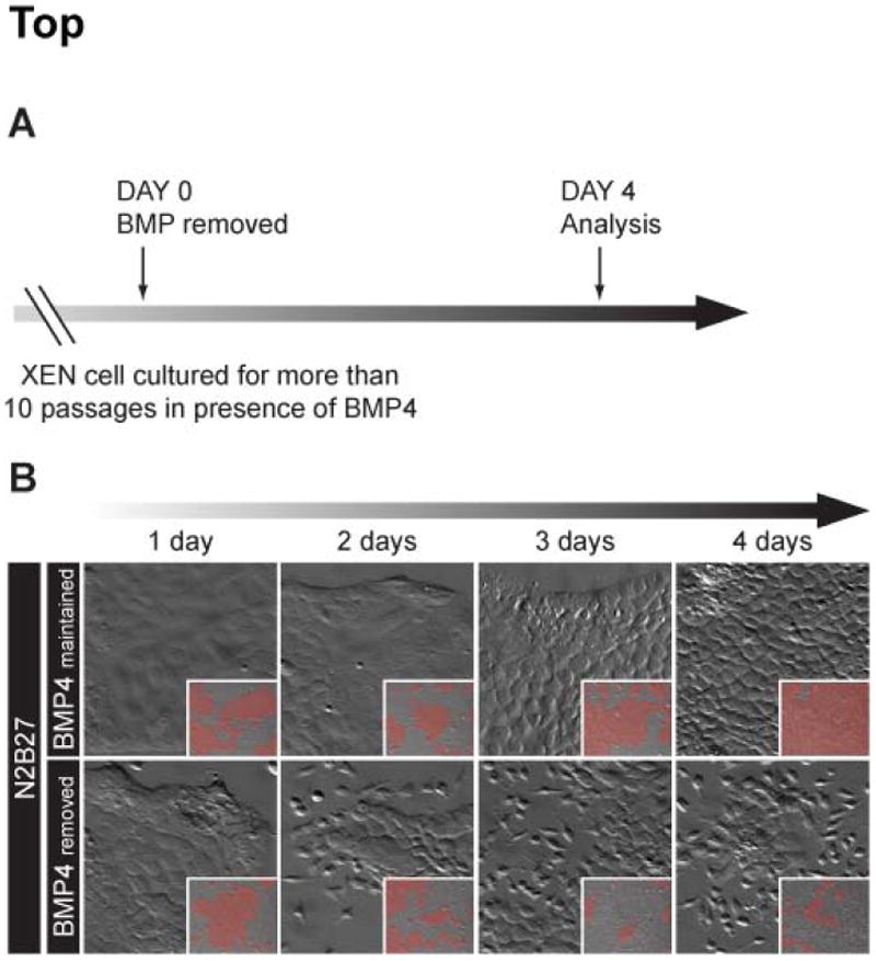
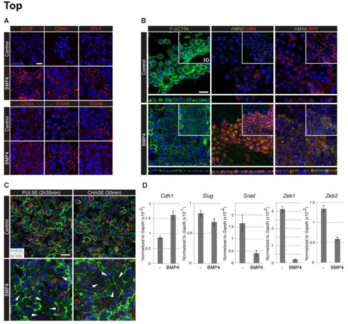
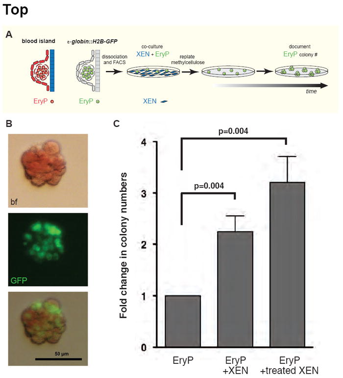
The visceral endoderm (VE) is an epithelial tissue in the early postimplantation mouse embryo that encapsulates the pluripotent epiblast distally and the extraembryonic ectoderm proximally. In addition to facilitating nutrient exchange before the establishment of a circulation, the VE is critical for patterning the epiblast. Since VE is derived from the primitive endoderm (PrE) of the blastocyst, and PrE-derived eXtraembryonic ENdoderm (XEN) cells can be propagated in vitro, XEN cells should provide an important tool for identifying factors that direct VE differentiation. In this study, we demonstrated that BMP4 signaling induces the formation of a polarized epithelium in XEN cells. This morphological transition was reversible, and was associated with the acquisition of a molecular signature comparable to extraembryonic (ex) VE. Resembling exVE which will form the endoderm of the visceral yolk sac, BMP4-treated XEN cells regulated hematopoiesis by stimulating the expansion of primitive erythroid progenitors. We also observed that LIF exerted an antagonistic effect on BMP4-induced XEN cell differentiation, thereby impacting the extrinsic conditions used for the isolation and maintenance of XEN cells in an undifferentiated state. Taken together, our data suggest that XEN cells can be differentiated towards an exVE identity upon BMP4 stimulation and therefore represent a valuable tool for investigating PrE lineage differentiation.


Figures

Figure 1
Figure 1. BMP treatment alters XEN cell morphology
(A) Schematic representation of periimplantation (E4.5) and early postimplantation (E5.5 and E7.25) stage mouse embryos. High magnification insets on the region where the exVE and extraembryonic mesoderm are apposed, representing the site of blood island formation (Ferkowicz and Yoder, 2005; Li et al., 2005). Red, EPI and its derivatives; green, TE and TE derivatives; blue, PrE and PrE derivatives. (B) Diagramatic representation of the timeline of BMP treatment. (C) XEN cell morphology after addition of BMP2 and BMP4 for 4 days at concentrations ranging from 5 to 20 ng/mL in serum culture conditions. (D) Kinetics of the morphological changes upon addition of 10 ng/mL of BMP4 in serum and serum-free (N2B27) conditions. (C-D) High magnification zooms of low magnification images (inset) acquired with a 10X magnification. In the insets, areas of epithelial cell colonies are highlighted in red.
Figure 2
Figure 2. Removal of BMP4 leads to a reversion of the morphological transition
(A) Diagramatic representation of BMP withdrawal scheme. (B) Kinetics of the morphological changes induced upon BMP4 removal (lower panels) compared to XEN cells maintained in serum-free (N2B27) medium supplemented with 10 ng/mL BMP4 (upper panels). High magnification zooms of low magnification images (inset) acquired with a 10X magnification. In the insets, areas of epithelial cell colonies are highlighted in red.
Figure 3
Figure 3. BMP4 stimulation induces the formation of a polarized epithelium
(A) Visualization of adherens junction (βCAT, CDH1), tight junction (ZO-1) and gap junction (CX43) constituent proteins in control and BMP4-treated XEN cells. BMP4 treatment also affected the localization and expression of Integrin α5 (ITGA5) and Integrin α6 (ITGA6) proteins. (B) Changes in the localization of F-actin, Amnionless (AMN), Cubilin (CUBN) and LRP2 proteins upon BMP4 treatment. Images correspond to single optical sections and orthogonal views (below), insets are 3D projections. (C) DiI-HDL uptake of control and BMP-4 treated XEN cells incubated for 2h 30min in presence (PULSE) and then for 30 min in absence (CHASE) of the dye. Arrowheads indicate cells with low levels of DiI-HDL. (D) qPCR analysis of MET transcriptional regulator gene expression in control (-) and BMP4-treated XEN cells. (A-C) βCAT, CDH1, ZO-1, CUBN, LRP2, Di-HDL, red; F-actin, AMN, green; nuclei counterstained with Hoechst, blue. Scale bars: 20 μm.
Figure 4
Figure 4. LIF signalling antagonizes the effect of BMP4
(A) Effect of BMP4 treatment on XEN cell morphology in absence (control) or in presence (LIF) of LIF. XEN cells were cultured for 4 days in serum conditions supplemented with 5 to 10 ng/mL BMP4 and 103 units/mL LIF. (B) Schematic representation of signal transduction pathways activated upon LIF ligand binding to LIFR/GP130 receptor and inhibitors used to block their activities. (C) Inhibition of P38 and JAK/STAT attenuates the inhibitory effect of LIF signalling on BMP driven MET. (A, C) Zoom magnified pictures of low magnification acquisitions (inset) acquired with a 10X zoom. In the insets, epithelial cell colonies were highlighted in red.
Figure 5
Figure 5. Microarray data analysis
(A) Diagramatic representation of the experimental scheme used for global gene expression analysis. Cells were treated for 10 days in presence of 25 ng/mL BMP4, a concentration optimal for the induction of the Afp∷GFP transgene. (B) Functional gene ontology analysis of the differentially expressed genes using the DAVID tool with a p-value ≤10-3. Top 50 genes downregulated (C) and upregulated (D) upon BMP4 treatment with their associated fold changes.
Figure 6
Figure 6. Fluctuation of Afp∷GFP reporter within the BMP-treated XEN cell population
(A) Detection of the Afp∷GFP transgene in control and BMP4-treated XEN cells. (B) percentage GFP-positive cells determined by flow cytometry upon culture for 4 days in the presence of various concentrations of BMP2 or BMP4 in serum and serum-free conditions. (C) Diagramatic representation of the experimental scheme used for global gene expression profiling in GFP+ and GFP- XEN cells maintained in 10 ng/mL BMP4. (D) The 5 genes found differentially upregulated (green, FC≥2) and downregulated (red, FC≤-2) in the GFP-positive population. (E) Kinetics of Afp∷GFP reporter expression after FACS sorting of the GFP-negative cells (upper panels) and the GFP-positive cells (lower panels). The diagrams depicts the evolution of the percentage of GFP-positive cells in the GFP- and GFP+ FACS sorted cells. (F) Single frames from 3D time-lapse imaging of Afp∷GFP Tg/+ XEN cells treated with 10 ng/mL BMP4. Annotated movie sequence is provided as Movie 1. Some cells upregulating GFP during the time-course are highlighted (red dashed lines depict cell perimeters). (A, E, F) GFP, green; nuclei counterstained with Hoechst, blue, brightfield, bf. Scale bars: 50 μm.
Figure 7
Figure 7. XEN cells promote EryP progenitor expansion
(A) Schematic representation of the assay used to test XEN cell ability to promote EryP expansion. (B) A representative red EryP colony expressing the ε-globin∷H2B-GFP transgene (GFP). Brigthfield, bf. Scale bar: 50μm. (C) The diagram depicts the average fold change in EryP colony number for EryP co-cocultured with XEN cells and BMP4-treated XEN cells compared to culture of EryP alone (n=6 experiments). P-values calculated from Mann-Whitney test indicate statistical significance.

References

    1. Anderson WJ, Zhou Q, Alcalde V, Kaneko OF, Blank LJ, Sherwood RI, Guseh JS, Rajagopal J, Melton DA. Genetic targeting of the endoderm with claudin-6CreER. Dev Dyn. 2008;237:504–12. - PMC - PubMed
    1. Arnold SJ, Robertson EJ. Making a commitment: cell lineage allocation and axis patterning in the early mouse embryo. Nat Rev Mol Cell Biol. 2009;10:91–103. - PubMed
    1. Artus J, Panthier JJ, Hadjantonakis AK. A role for PDGF signaling in expansion of the extra-embryonic endoderm lineage of the mouse blastocyst. Development. 2010;137:3361–72. - PMC - PubMed
    1. Artus J, Piliszek A, Hadjantonakis AK. The primitive endoderm lineage of the mouse blastocyst: Sequential transcription factor activation and regulation of differentiation by Sox17. Dev Biol. 2011;350:393–404. - PMC - PubMed
    1. Artus J, Vandormael-Pournin S, Frodin M, Nacerddine K, Babinet C, Cohen-Tannoudji M. Impaired mitotic progression and preimplantation lethality in mice lacking OMCG1, a new evolutionarily conserved nuclear protein. Mol Cell Biol. 2005;25:6289–302. - PMC - PubMed

Publication types

MeSH terms

Substances