. 2011 Nov 3:11:476.
doi: 10.1186/1471-2407-11-476.

GnRH receptor activation competes at a low level with growth signaling in stably transfected human breast cell lines

Affiliations

GnRH receptor activation competes at a low level with growth signaling in stably transfected human breast cell lines

Kevin Morgan et al. BMC Cancer. .

Abstract

Background: Gonadotrophin releasing hormone (GnRH) analogs lower estrogen levels in pre-menopausal breast cancer patients. GnRH receptor (GnRH-R) activation also directly inhibits the growth of certain cells. The applicability of GnRH anti-proliferation to breast cancer was therefore analyzed.

Methods: GnRH-R expression in 298 primary breast cancer samples was measured by quantitative immunofluorescence. Levels of functional GnRH-R in breast-derived cell lines were assessed using 125I-ligand binding and stimulation of 3H-inositol phosphate production. Elevated levels of GnRH-R were stably expressed in cells by transfection. Effects of receptor activation on in vitro cell growth were investigated in comparison with IGF-I and EGF receptor inhibition, and correlated with intracellular signaling using western blotting.

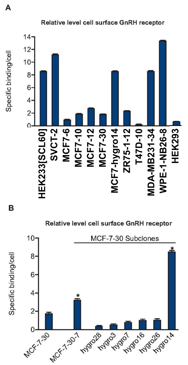
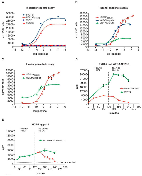
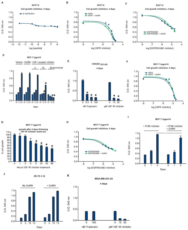
Results: GnRH-R immunoscoring was highest in hormone receptor (triple) negative and grade 3 breast tumors. However prior to transfection, functional endogenous GnRH-R were undetectable in four commonly studied breast cancer cell lines (MCF-7, ZR-75-1, T47D and MDA-MB-231). After transfection with GnRH-R, high levels of cell surface GnRH-R were detected in SVCT and MDA-MB-231 clones while low-moderate levels of GnRH-R occurred in MCF-7 clones and ZR-75-1 clones. MCF-7 sub-clones with high levels of GnRH-R were isolated following hygromycin phosphotransferase transfection. High level cell surface GnRH-R enabled induction of high levels of 3H-inositol phosphate and modest growth-inhibition in SVCT cells. In contrast, growth of MCF-7, ZR-75-1 or MDA-MB-231 clones was unaffected by GnRH-R activation. Cell growth was inhibited by IGF-I or EGF receptor inhibitors. IGF-I receptor inhibitor lowered levels of p-ERK1/2 in MCF-7 clones. Washout of IGF-I receptor inhibitor resulted in transient hyper-elevation of p-ERK1/2, but co-addition of GnRH-R agonist did not alter the dynamics of ERK1/2 re-phosphorylation.

Conclusions: Breast cancers exhibit a range of GnRH-R immunostaining, with higher levels of expression found in triple-negative and grade 3 cancers. However, functional cell surface receptors are rare in cultured cells. Intense GnRH-R signaling in transfected breast cancer cells did not markedly inhibit growth, in contrast to transfected HEK 293 cells indicating the importance of intracellular context. GnRH-R signaling could not counteract IGF-I receptor-tyrosine kinase addiction in MCF-7 cells. These results suggest that combinatorial strategies with growth factor inhibitors will be needed to enhance GnRH anti-proliferative effects in breast cancer.


Figures

Figure 1
Figure 1
GnRH receptor is expressed across a wide range in breast cancer and is highest in triple negative tumours when measured by immunostaining. Association of GnRH receptor expression and (A) Cancer type and (B) Cancer grade. Quantitative immunofluorescence (AQUA) was used to measure GnRH receptor. One way ANOVA was used to test for significant differences between subtypes, the mean for each group is shown with a dashed line. C. Representative examples of high GnRH receptor expression (top images) and low expression (bottom images). Left hand images are immunohistochemical images of tissue microarray (TMA) cores of individual breast cancer with brown staining corresponding to GnRH receptor expression and blue to haematoxylin staining. Right hand images are immunofluorescence images of TMA cores, with red staining corresponding to GnRH receptor expression, blue (DAPI) staining indicating cell nuclei and green staining detecting cytokeration (ie carcinoma cell) staining. White arrows indicate areas of positive expression.
Figure 2
Figure 2
Stably transfected breast cell lines can be generated with functional GnRH receptor. Relative levels of GnRH at the cell surface detected by ligand binding assay in human cell lines stably transfected with rat GnRH receptor cDNA expression construct A. Subclones of MCF-7 clone 30 expressing modified levels of GnRH receptor at the cell surface were isolated. HEK293 and T47-D10 cells demonstrated background levels of binding. All other cell lines shown demonstrated significantly (p < 0.05 ANOVA) higher levels of specific binding. B. MCF-7-30-7 was subcloned from MCF-7-30 and then transfected with a PvuII SV40-hygromycin resistance gene fragment. Clones resistant to G418 and hygromycin were screened for altered GnRH receptor expression. GnRH receptor levels were elevated in clone MCF-7-30-hygro14, similar to levels in HEK293[SCL60]. * p < 0.05 (ANOVA followed by Dunnett's test) indicates significantly higher in MCF-7-30-7 and MCF-7-30-hygro14 relative to MCF-7-30 binding.
Figure 3
Figure 3
Dynamics of GnRH receptor activation following treatment with Triptorelin. Treatment of stably transfected cells with GnRH elicited high levels of 3H- inositol phosphate (IP) production (A-C). Removal of GnRH and LiCl revealed the dynamics of 3H- inositol phosphate turnover in different cell types (D,E). The decrease in levels of 3H- inositol phosphates was slower in SVCT-2 cells. Statistically different values (p < 0.05 ANOVA followed by Dunnett's test) compared to control values were as follows; for A, All values shown for SVCT-2 and HEK293[SCL60] cells > -10 log [peptide]; for B, all values shown > -10 log [peptide] for all 3 cell lines; for C, all values shown > -10 log [peptide] for both cell lines; for D, all values shown for SVCT-2 and all values up to 180 min for WPE-1-NB26-8; for E, all values from 30 min to 180 min.
Figure 4
Figure 4
The effect of Triptorelin on growth of cells stably transfected with GnRH. A. Growth of SVCT-2 after 4 days was marginally inhibited (10-18%) by treatment with Triptorelin. However, cell growth was effectively inhibited by IGF-IR inhibitor II and co-treatment with Triptorelin exerted a small additive effect (B). EGFR/ErbB2 inhibitor reduced SVCT-2 cell growth, but co-treatment with Triptorelin had no effect (C). Growth of MCF-7-30-7hygro14 cells was not affected by treatment with 100 nM Triptorelin (D), unlike HEK293[SCL60] cells (E) after 4 days. Growth and survival were inhibited by IGF-IR inhibitor II but co-treatment with Triptorelin had no effect (F). Transient exposure to 15 μM IGF-IR inhibitor II for up to 2 hours resulted in less than 10% growth-inhibition after 4 days, longer exposures resulted in more extensive growth-inhibition (G). Growth of MCF-7-30-7hygro14 cells was inhibited by EGFR/ErbB2 inhibitor but not affected by treatment with 7 μM PI3Kγ inhibitor and co-treatment with 100 nM Triptorelin exerted no significant growth-inhibition (H and I). Growth of ZR75-1-12 (J) and MDA-MB-231-34 (K) was unaffected by treatment with 100 nM Triptorelin. * p < 0.05 (ANOVA followed by Dunnett's test).
Figure 5
Figure 5
The level of p-ERK1/2 is influenced by integration of signaling from multiple cell surface receptors, blocking the response to activated GnRH receptor. A. Triptorelin did not affect levels of p-ERK1/2 in serum-starved MCF-7-30 or MDA-MB-231-34 cells. B. Treatment of stably transfected cells with 100 nM Triptorelin transiently elevated levels of phosphorylated ERK1/2 (p-ERK1/2) in serum-starved MCF-7-30-7hygro14 cells but not in the presence of serum. Bar graphs indicate effect of no serum vs with serum on ERK response in MCF7hygro14, statistically significant for no serum, p < 0.05. C. Treatment with IGF-IR inhibitor resulted in rapid and sustained de-phosphorylation of ERK1/2 in MCF-7-30-7hygro14 cells but not in MDA-MB-231-34 cells. D. Rapid re-phosphorylation of ERK1/2 occurred in MCF-7-30-7hygro14 cells when IGF-I receptor inhibitor was washed off and replaced with fresh culture medium but addition of 100 nM Triptorelin did not affect levels of phosphorylated ERK1/2 Re-phosphorylation of ERK1/2 was less marked in HEK293[SCL60] cells and addition of Triptorelin considerably augmented levels of phosphorylated ERK1/2.

Similar articles

Cited by

References

    1. LHRH-agonists in Early Breast Cancer Overview group. Cuzick J, Ambroisine L, Davidson N, Jakesz R, Kaufmann M, Regan M, Sainsbury R. Use of luteinising-hormone-releasing hormone agonists as adjuvant treatment in premenopausal patients with hormone-receptor-positive breast cancer: a meta-analysis of individual patient data from randomised adjuvant trials. Lancet. 2007;369:1711–2173. - PubMed
    1. Gnant M, Mlineritsch B, Schippinger W, Luschin-Ebengreuth G, Pöstlberger S, Menzel C, Jakesz R, Seifert M, Hubalek M, Bjelic-Radisic V, Samonigg H, Tausch C, Eidtmann H, Steger G, Kwasny W, Dubsky P, Fridrik M, Fitzal F, Stierer M, Rücklinger E, Greil R. ABCSG-12 Trial Investigators. Marth C. Endocrine therapy plus zoledronic acid in premenopausal breast cancer. New Engl J Med. 2009;360:679–691. doi: 10.1056/NEJMoa0806285. - DOI - PubMed
    1. Segal-Abramson T, Kitroser H, Levy J, Schally AV, Sharoni Y. Direct effects of luteinizing hormone-releasing hormone agonists and antagonists on MCF-7 mammary cancer cells. Proc Natl Acad Sci USA. 1992;89:2336–2339. doi: 10.1073/pnas.89.6.2336. - DOI - PMC - PubMed
    1. Hershkovitz E, Marbach M, Bosin E, Levy J, Roberts CT Jr, LeRoith D, Schally AV, Sharoni Y. Luteinizing hormone-releasing hormone antagonists interfere with autocrine and paracrine growth stimulation of MCF-7 mammary cancer cells by insulin-like growth factors. J Clin Endocrinol Metab. 1993;77:963–968. doi: 10.1210/jc.77.4.963. - DOI - PubMed
    1. Buchholz S, Seitz S, Schally AV, Engel JB, Rick FG, Szalontay L, Hohla F, Krishan A, Papadia A, Gaiser T, Brockhoff G, Ortmann O, Diedrich K, Köster F. Triple-negative breast cancers express receptors for luteinizing hormone-releasing hormone (LHRH) and respond to LHRH antagonist cetrorelix with growth inhibition. Int J Oncol. 2009;35:789–796. - PubMed

Publication types