

Inherited liver shunts in dogs elucidate pathways regulating embryonic development and clinical disorders of the portal vein
- PMID: 22052005
- PMCID: PMC3275728
- DOI: 10.1007/s00335-011-9364-0
Inherited liver shunts in dogs elucidate pathways regulating embryonic development and clinical disorders of the portal vein
Abstract
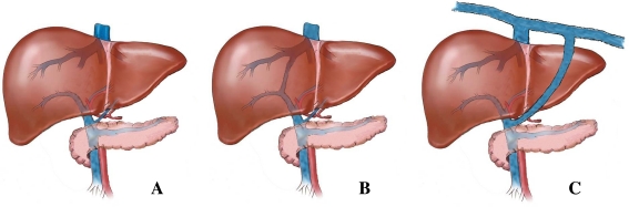
Congenital disorders of the hepatic portal vasculature are rare in man but occur frequently in certain dog breeds. In dogs, there are two main subtypes: intrahepatic portosystemic shunts, which are considered to stem from defective closure of the embryonic ductus venosus, and extrahepatic shunts, which connect the splanchnic vascular system with the vena cava or vena azygos. Both subtypes result in nearly complete bypass of the liver by the portal blood flow. In both subtypes the development of the smaller branches of the portal vein tree in the liver is impaired and terminal branches delivering portal blood to the liver lobules are often lacking. The clinical signs are due to poor liver growth, development, and function. Patency of the ductus venosus seems to be a digenic trait in Irish wolfhounds, whereas Cairn terriers with extrahepatic portosystemic shunts display a more complex inheritance. The genes involved in these disorders cannot be identified with the sporadic human cases, but in dogs, the genome-wide study of the extrahepatic form is at an advanced stage. The canine disease may lead to the identification of novel genes and pathways cooperating in growth and development of the hepatic portal vein tree. The same pathways likely regulate the development of the vascular system of regenerating livers during liver diseases such as hepatitis and cirrhosis. Therefore, the identification of these molecular pathways may provide a basis for future proregenerative intervention.
Figures
References
-
- Abernethy J. Account of two instances of uncommon formation in the viscera of the human body. Philos Trans R Soc Lond B Biol Sci. 1793;83:295–299.
-
- Adeagbo AS, Breen CA, Cutz E, Lees JG, Olley PM, Coceani F. Lamb ductus venosus: evidence of a cytochrome P-450 mechanism in its contractile tension. J Pharmacol Exp Ther. 1990;252:875–879. - PubMed
-
- Allen L, Stobie D, Mauldin GN, Baer KE. Clinicopathologic features of dogs with hepatic microvascular dysplasia with and without portosystemic shunts: 42 cases (1991–1996) J Am Vet Med Assoc. 1999;214:218–220. - PubMed
Publication types
MeSH terms
Substances
LinkOut - more resources
Full Text Sources
