. 2012 Feb 1;302(3):G326-35.
doi: 10.1152/ajpgi.00083.2011. Epub 2011 Nov 3.

Resveratrol causes cell cycle arrest, decreased collagen synthesis, and apoptosis in rat intestinal smooth muscle cells

Affiliations

Resveratrol causes cell cycle arrest, decreased collagen synthesis, and apoptosis in rat intestinal smooth muscle cells

Patricia Garcia et al. Am J Physiol Gastrointest Liver Physiol. .

Abstract

One of the most difficult and treatment-resistant complications of Crohn's disease is the development of fibrotic intestinal strictures due to mesenchymal cell hyperplasia and collagen deposition. Resveratrol, a phytoalexin found in berries, peanuts, grapes, and red wine, has been shown to inhibit fibrosis in vasculature, heart, lung, kidney, liver, and esophagus in animal models. Resveratrol has also been shown to inhibit oxidation, inflammation, and cell proliferation and to decrease collagen synthesis in several cell types or animal models. The aim of this study was to determine whether resveratrol has antifibrotic effects on intestinal smooth muscle cells. Responses to resveratrol by cultured smooth muscle cells isolated from colons of untreated Lewis rats were examined; this rat strain is used in a model of Crohn's disease with prominent intestinal fibrosis. A relative decrease in cell numbers following treatment with 50 and 100 μM resveratrol was evident at 24 h (P ≤ 0.005). This effect was largely due to cell cycle arrest, with an increase in the percent of cells in S phase from 8 to 25-35% (P < 0.05). Cell viability was unchanged until 2-3 days of treatment when there was a 1.2- to 5.0-fold increase in the percent of apoptotic cells, depending on the assay (P < 0.05). Expression of collagen type I protein was decreased following treatment with resveratrol for 24 h (to 44 and 25% of control levels with 50 and 100 μM resveratrol, respectively; P < 0.05). Expression of procollagen types I and III mRNA was also decreased with resveratrol treatment. Resveratrol (50 μM) diminished the proliferative response to TGF-β₁ (P = 0.02) as well as IGF-I-stimulated collagen production (P = 0.02). Thus resveratrol decreases intestinal smooth muscle cell numbers through its effects on cell cycle arrest and apoptosis and also decreases collagen synthesis by the cells. These effects could be useful in preventing the smooth muscle cell hyperplasia and collagen deposition that characterize stricture formation in Crohn's disease.


Figures
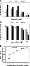
Fig. 1.
Fig. 1.
A: effect of resveratrol (Res) on proliferation of rat intestinal smooth muscle cells (RISM). All treatments were begun on day 0 and continued for 1, 3, or 5 days. At the end of treatment, cells were trypsinized, stained with Trypan blue, and counted in a hemacytometer. Means of counts of triplicate wells are expressed relative to that of untreated cells from cultures counted on day 0 and are the results of an experiment representative of 2. Error bars represent SD. Statistical comparisons are between treated and vehicle-exposed cultures from the same day (*P < 0.05; **P < 0.005; ***P < 0.0005). B: mitochondrial dehydrogenase (MDH) activity. Mean background-corrected A450s of triplicate wells are expressed relative to that of vehicle-exposed cells from the same day. Results shown are from parallel cultures from the same representative (of 2) experiment as shown in A above. Error bars represent SD. Statistical comparisons are between treated and vehicle-exposed cultures from the same day (*P < 0.05; **P < 0.005; ***P < 0.0005). C: correlation of relative MDH activity with relative cell numbers. Data are from A and B above, but here the cell numbers are expressed relative to those of vehicle-exposed cultures of the same day (“x” symbol, DMSO; circles, Res 1 μM; triangles, Res 10 μM; diamonds, Res 50 μM; squares, Res 100 μM; open symbols, day 1; shaded symbols, day 3; solid symbols, day 5). The outlier at bottom left (arrow) represents data for cultures treated with 100 μM Res for 5 days.
Fig. 2.
Fig. 2.
Effect of resveratrol on cell cycle in RISM. Cultures of RISM were treated with varying concentrations of resveratrol for 24 h. At the end of treatment, cells were trypsinized, incubated with DNase-free RNase, stained with propidium iodide (PI), and analyzed by flow cytometry. Peak integration was done using ModFitLT V3.1 (PMac) software. Results shown are of an experiment representative of 4, each with duplicate or triplicate cultures.
Fig. 3.
Fig. 3.
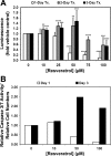
A: effect of resveratrol on caspase-3/7 activation in RISM. Cultures of RISM were treated with varying concentrations of resveratrol for 1, 2, or 3 days. At the end of treatment (Tx), cells were lysed in the culture wells and a caspase-3/7 substrate was applied. After 1 h incubation at room temperature, the amount of the luminescent product generated was measured. Values are means of 4–11 experiments, each with duplicate wells. Error bars represent SD. Statistical comparisons are between treated and vehicle-exposed cultures from the same day (**P ≤ 0.005; ***P ≤ 0.0005; ****P ≤ 0.00005). B: ratio of caspase-3/7 activation in 3-day resveratrol-treated cultures from 5–7 of the experiments shown in A (i.e., those experiments that contained the relevant data points; luminescence expressed as fold vehicle control) to mean cell counts from 3-day treated cultures (mean of Fig. 1A data plus data from the replicate experiment; expressed here as fold vehicle control) plotted against resveratrol concentration.
Fig. 4.
Fig. 4.
Effect of resveratrol on exposure of phosphatidylserine on the outer surface of the plasma membrane in RISM. A: cultures of RISM were treated with 50 or 100 μM resveratrol for 3 days. At the end of treatment, the cells were trypsinized, incubated with FITC-labeled annexin V and propidium iodide, and analyzed by flow cytometry. Representative quadrant graphs are shown. B: numbers of FITC-positive propidium iodide-negative cells were expressed as the percent of total gated cells. The data were then expressed as fold vehicle control. Error bars represent SD. Statistical comparisons are between treated and vehicle-exposed cells (*P < 0.05; ***P < 0.00005). Results shown are of an experiment representative of 2, each with duplicate cultures. C: cultures of RISM were treated with 100 μM resveratrol for 2 days. Cells incubated with 0.5 μM staurosporine (Stauro) for 6.5 h served as positive controls. Without trypsinization, the treated cells were incubated with FITC-annexin V and propidium iodide and examined by fluorescence microscopy. Shown are representative low-power microscopic fields from an experiment with duplicate wells.
Fig. 5.
Fig. 5.
Effect of resveratrol on DNA fragmentation in RISM. Cultures of RISM were treated with varying concentrations of resveratrol for 2 days. At the end of treatment, a terminal deoxynucleotidyl transferase-mediated dUTP nick-end labeling (TUNEL) assay was done. The cells were trypsinized, nick end-labeled with bromodeoxyuridine (BrdU), which was then detected by use of a FITC-labeled antibody, stained with propidium iodide, and analyzed by flow cytometry. A: representative quadrant graphs are shown. B: double-stained cells were counted and expressed as the percent of total gated cells. The data were then expressed as fold vehicle control. Error bars represent SD. Statistical comparisons are between treated and vehicle-exposed cells (*P < 0.05). Results shown are means of 2 experiments, each with duplicate or quadruplicate cultures.
Fig. 6.
Fig. 6.
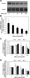
Effect of resveratrol on collagen expression in RISM. Cultures of RISM were treated with varying concentrations of resveratrol for 1 day. A: at the end of treatment, the cells were lysed and subjected to nonreducing SDS-PAGE. Blots were probed with an antibody to collagen type I and subsequently with an antibody to GAPDH. The blot shown is representative of 2 experiments, each with duplicate cultures. B: densitometric quantification of the specific collagen type I doublet bands normalized to the total GAPDH immunoreactivity. These quantitative data represent the means of duplicate gels from both of the experiments. C and D: cells from parallel cultures treated for 1 day and cultures from separate experiments treated for 2 or 3 days were analyzed by quantitative real-time PCR. Each bar represents the means of 2 experiments, each with duplicate wells. C: results for procollagen type I. D: results for procollagen type III. Error bars represent SD. Statistical comparisons are between treated and vehicle-exposed cells (*P < 0.05; **P < 0.005).
Fig. 7.
Fig. 7.
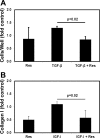
Effect of resveratrol on cytokine stimulation of cultured RISM. Cultures of RISM were transitioned to serum-free medium. Twenty-four hours later, fibrogenic cytokines were introduced concurrently with resveratrol (50 μM) or vehicle. A: effect of resveratrol on TGF-β1-induced proliferation. At the end of treatment (1–3 days; 1 ng/ml TGF-β1) cells were trypsinized, stained with Trypan blue, and counted in a hemacytometer. Mean counts of 4–6 wells/condition were calculated and expressed relative to that of vehicle exposed cultures. Results shown represent the means of 3 experiments. Error bars represent SD. B: effect of resveratrol on IGF-I-induced collagen type I protein expression. At the end of treatment (2–3 days; 100 ng/ml IGF-I), the cells were lysed and subjected to nonreducing SDS-PAGE. Blots were probed with an antibody to collagen type I and subsequently with an antibody to GAPDH. Densitometric quantification of collagen type I immunoreactivity relative to that of GAPDH is shown. The data represent the means of 2 experiments, each with duplicate cultures. Error bars represent SD.

Similar articles

Cited by

References

    1. Almeida L, Vaz-da-Silva M, Falcão A, Soares E, Costa R, Loureiro AI, Fernandes-Lopes C, Rocha J-F, Nunes T, Wright L, Soares-da-Silva P. Pharmacokinetic and safety profile of trans-resveratrol in a rising multiple-dose study in healthy volunteers. Mol Nutr Food Res 53: S7–S15, 2009 - PubMed
    1. Baeuerle PA, Henkel T. Function and activation of NF-κB in the immune system. Annu Rev Immunol 12: 141–179, 1994 - PubMed
    1. Banerejee S, Bueso-Ramos C, Aggarwal BB. Suppression of 7,12-dimethylbenz(a)anthracene-induced mammary carcinogenesis in rats by resveratrol: role of nuclear factor-κB, cyclooxygenase 2, and matrix metalloprotease 9. Cancer Res 62: 4945–4954, 2002 - PubMed
    1. Baur JA, Sinclair DA. Therapeutic potential of resveratrol: the in vivo evidence. Nat Rev Drug Discov 5: 493–506, 2006 - PubMed
    1. Boocock DJ, Faust GES, Patel KR, Schinas AM, Brown VA, Ducharme MP, Booth TD, Crowell JA, Perloff M, Gescher AJ, Steward WP, Brenner DE. Phase I dose escalation pharmacokinetic study in healthy volunteers of resveratrol, a potential cancer chemopreventive agent. Cancer Epidemiol Biomarkers Prev 16: 1246–1252, 2007 - PubMed

Publication types

MeSH terms

LinkOut - more resources