. 2011 Dec 15;71(24):7360-5.
doi: 10.1158/0008-5472.CAN-11-2023. Epub 2011 Nov 3.

5-Hydroxymethylcytosine is strongly depleted in human cancers but its levels do not correlate with IDH1 mutations

Affiliations

5-Hydroxymethylcytosine is strongly depleted in human cancers but its levels do not correlate with IDH1 mutations

Seung-Gi Jin et al. Cancer Res. .

Abstract

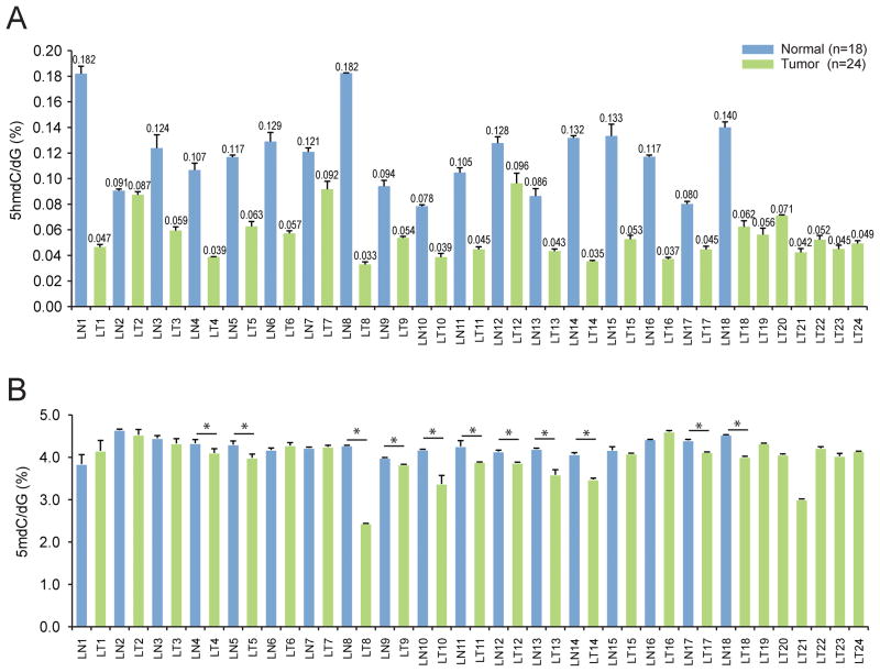
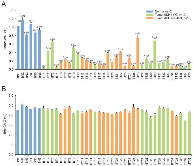
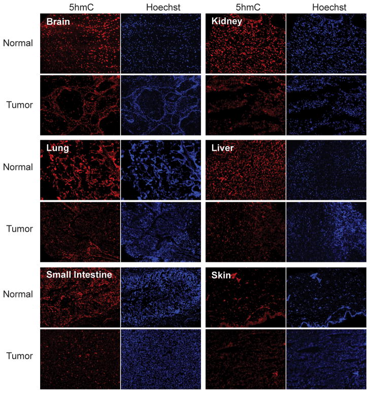
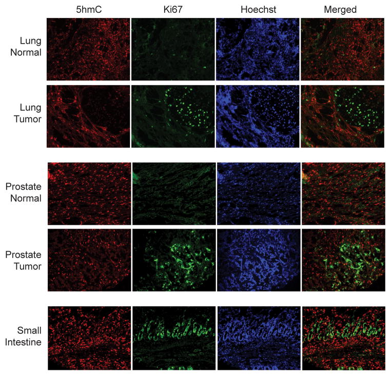
The base 5-hydroxymethylcytosine (5hmC) was recently identified as an oxidation product of 5-methylcytosine in mammalian DNA. Here, using sensitive and quantitative methods to assess levels of 5-hydroxymethyl-2'-deoxycytidine (5hmdC) and 5-methyl-2'-deoxycytidine (5mdC) in genomic DNA, we investigated whether levels of 5hmC can distinguish normal tissue from tumor tissue. In squamous cell lung cancers, levels of 5hmdC were depleted substantially with up to 5-fold reduction compared with normal lung tissue. In brain tumors, 5hmdC showed an even more drastic reduction with levels up to more than 30-fold lower than in normal brain, but 5hmdC levels were independent of mutations in isocitrate dehydrogenase-1. Furthermore, immunohistochemical analysis indicated that 5hmC is remarkably depleted in many types of human cancer. Importantly, an inverse relationship between 5hmC levels and cell proliferation was observed with lack of 5hmC in proliferating cells. The data therefore suggest that 5hmdC is strongly depleted in human malignant tumors, a finding that adds another layer of complexity to the aberrant epigenome found in cancer tissue. In addition, a lack of 5hmC may become a useful biomarker for cancer diagnosis.


Figures

Figure 1
Figure 1. Quantitation of 5hmdC and 5mdC in normal lung and lung squamous cell carcinoma DNA
A. 5hmdC. The first 18 samples are matched normal lung (LN, blue) and lung tumors (LT, green). The last six samples are lung tumors without available normal tissue. B. 5mdC. The asterisks indicate that the levels of 5mdC were significantly reduced in the tumor compared to normal lung (P < 0.05).
Figure 2
Figure 2. Quantitation of 5hmdC and 5mdC in normal brain DNA and in stage II/III astrocytomas
A. 5hmdC quantitation in normal brain (NB, blue) and in brain tumors (BT, green or orange). Samples BT1–16, BT25, BT27–29 and BT32–36 were stage III astrocytomas; BT17–24, BT26, BT30 and BT31 were stage II astrocytomas, and BT37 and BT38 were glioblastomas. B. 5mdC in normal brain (NB) and in brain tumors (BT). Tumors with no IDH1 mutation are shown in green; tumors withIDH1 R132H are shown in orange. The sample BT26 had a minor allele frequency of IDH1 R132H. Sample BT25 had the rare mutation R132G.
Figure 3
Figure 3. Immunohistochemical analysis of 5hmC on human tissue arrays
Human tissue arrays containing samples of malignant tumor and corresponding normal tissue were stained with anti-5hmC antibody. Staining with Hoechst 33258 is shown as a control. The magnification of all panels is 10-fold.
Figure 4
Figure 4. Inverse relationship between 5hmC and Ki67 staining
Sections of normal lung and lung tumor, normal prostate and prostate tumor and normal small intestine were stained with anti-5hmC (red) and anti-Ki67 antibodies (green) to mark proliferating cells. Note the mutually exclusive staining of 5hmC and Ki67 in the tissue sections. For example, proliferating cells in intestinal crypts are positive for Ki67 but negative for 5hmC.

References

    1. Kriaucionis S, Heintz N. The nuclear DNA base 5-hydroxymethylcytosine is present in Purkinje neurons and the brain. Science. 2009;324:929–930. - PMC - PubMed
    1. Tahiliani M, Koh KP, Shen Y, Pastor WA, Bandukwala H, Brudno Y, et al. Conversion of 5-methylcytosine to 5-hydroxymethylcytosine in mammalian DNA by MLL partner TET1. Science. 2009;324:930–935. - PMC - PubMed
    1. Ito S, D’Alessio AC, Taranova OV, Hong K, Sowers LC, Zhang Y. Role of Tet proteins in 5mC to 5hmC conversion, ES-cell self-renewal and inner cell mass specification. Nature. 2010 in press. - PMC - PubMed
    1. Globisch D, Munzel M, Muller M, Michalakis S, Wagner M, Koch S, et al. Tissue distribution of 5-hydroxymethylcytosine and search for active demethylation intermediates. PLoS One. 2010;5:e15367. - PMC - PubMed
    1. Szwagierczak A, Bultmann S, Schmidt CS, Spada F, Leonhardt H. Sensitive enzymatic quantification of 5-hydroxymethylcytosine in genomic DNA. Nucleic Acids Res. 2010;38:e181. - PMC - PubMed

Publication types

MeSH terms