Review
. 2011 Nov;8(6):504-7.
doi: 10.1513/pats.201102-022MW.

Dynamics of airway blood vessels and lymphatics: lessons from development and inflammation

Affiliations
Review

Dynamics of airway blood vessels and lymphatics: lessons from development and inflammation

Donald M McDonald et al. Proc Am Thorac Soc. 2011 Nov.

Abstract

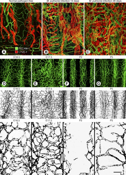
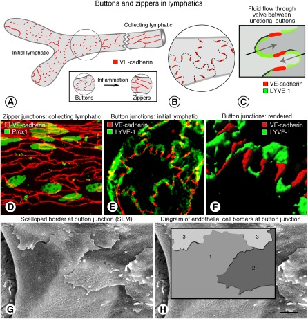
Blood vessels and lymphatic vessels in the respiratory tract play key roles in inflammation. By undergoing adaptive remodeling and growth, blood vessels undergo changes that enable the extravasation of plasma and leukocytes into inflamed tissues, and lymphatic vessels adjust to the increased fluid clearance and cell traffic involved in immune responses. Blood vessels and lymphatics in adult airways are strikingly different from those of late-stage embryos. Before birth, blood vessels in mouse airways make up a primitive plexus similar to that of the yolk sac. This plexus undergoes rapid and extensive remodeling at birth. In the early neonatal period, parts of the plexus regress. Capillaries then rapidly regrow, and with arterioles and venules form the characteristic adult vascular pattern. Lymphatic vessels of the airways also undergo rapid changes around birth, when lymphatic endothelial cells develop button-like intercellular junctions specialized for efficient fluid uptake. Among the mechanisms that underlie the onset of rapid vascular remodeling at birth, changes in tissue oxygen tension and mechanical forces associated with breathing are likely to be involved, along with growth factors that promote the growth and maturation of blood vessels and lymphatics. Whatever the mechanisms, the dynamic nature of airway blood vessels and lymphatics during perinatal development foretells the extraordinary vascular plasticity found in many diseases.


Figures

Figure 1.
Figure 1.
(AC) Remodeling of airway blood vessels and lymphatics in mice after Mycoplasma pulmonis infection. Confocal micrographs of mouse tracheal whole mounts stained for blood vessels (PECAM-1, green) and lymphatic vessels (LYVE-1, red). (A) Pathogen-free mouse. (B) Mouse with M. pulmonis infection of the respiratory tract for 14 days. (C) Mouse infected for 28 days. (DG) Pre- and postnatal changes in tracheal blood vessels from E16.5 through P5. Confocal micrographs of tracheal whole mounts with blood vessels stained for PECAM-1. (D, E) At E16.5 and E17.5 the tracheal vasculature is a primitive plexus of highly anastomotic, undifferentiated blood vessels. (F) At P2 the vasculature is segmented by pruning of the primitive plexus over cartilage rings. (G) At P5 the adult vascular pattern is evident after horizontal capillaries have grown over cartilage rings and arterioles and venules form between the rings. The second row of images shows PECAM-1 staining of blood vessels in color. The third row shows the same images in grayscale to emphasize the conspicuous differences in the vasculature patterns. The bottom row of images shows higher magnification views of the vascular architecture. Scale bar at bottom right is 100 μm in top row, 80 μm in second and third rows, and 20 μm in bottom row. (AC reprinted with permission from Reference ; DG reprinted with permission from Reference 28).
Figure 2.
Figure 2.
(AC) Buttons at sites of fluid entry in initial lymphatics in mice. (A) Schematic diagram showing distinctive, discontinuous buttons in endothelium of initial lymphatics and continuous zippers in collecting lymphatics as revealed by vascular endothelial (VE)-cadherin immunoreactivity. (B) More detailed diagram of the oak leaf–shaped endothelial cells (dashed lines) of initial lymphatics. Buttons (red) appear to be oriented perpendicular to the cell border but are in fact parallel to the sides of flaps. Most LYVE-1 immunoreactivity is at the tips of flaps. (C) Enlarged diagram of buttons showing complementary shapes of overlapping flaps of adjacent oak leaf–shaped endothelial cells. Adherens junctions at the sides of flaps are believed to direct fluid entry (arrows) through the junction-free region at the tip. (DH) Zipper and button junctions in endothelium of lymphatics. (D) Confocal image showing VE-cadherin immunoreactivity (red) at continuous zippers in collecting lymphatic identified by Prox1 (green) in nuclei. (E) Confocal image showing VE-cadherin at buttons (red) and LYVE-1 between buttons (green) at the border of oak leaf–shaped endothelial cells of initial lymphatic. (F) Rendered version of region in E shown here at higher magnification. (G) Scanning electron microscopic image showing external surface of overlapping flaps at the junction of three endothelial cells of initial lymphatic. (H) Drawing of region in G showing the contributions of three endothelial cells. Scale bar is 10 μm in DE, 3 μm in F, and 1.4 μm in GH. (Reprinted with permission from Reference 26).

References

    1. Dunnill MS. The pathology of asthma, with special reference to changes in the bronchial mucosa. J Clin Pathol 1960;13:27–33 - PMC - PubMed
    1. Wilson JW, Hii S. The importance of the airway microvasculature in asthma. Curr Opin Allergy Clin Immunol 2006;6:51–55 - PubMed
    1. Ebina M. Remodeling of airway walls in fatal asthmatics decreases lymphatic distribution: beyond thickening of airway smooth muscle layers. Allergol Int 2008;57:1–10 - PubMed
    1. Detoraki A, Granata F, Staibano S, Rossi FW, Marone G, Genovese A. Angiogenesis and lymphangiogenesis in bronchial asthma. Allergy 2010;65:946–958 - PubMed
    1. Ribatti D, Puxeddu I, Crivellato E, Nico B, Vacca A, Levi-Schaffer F. Angiogenesis in asthma. Clin Exp Allergy 2009;39:1815–1821 - PubMed

Publication types

LinkOut - more resources