. 2012 Jan;32(1):5-14.
doi: 10.1161/ATVBAHA.111.238725. Epub 2011 Nov 3.

Determinants of microvascular network topologies in implanted neovasculatures

Affiliations

Determinants of microvascular network topologies in implanted neovasculatures

Carlos C Chang et al. Arterioscler Thromb Vasc Biol. 2012 Jan.

Abstract

Objective: During neovascularization, the end result is a new functional microcirculation composed of a network of mature microvessels with specific topologies. Although much is known concerning the mechanisms underlying the initiation of angiogenesis, it remains unclear how the final architecture of microcirculatory beds is regulated. To begin to address this, we determined the impact of angiogenic neovessel prepatterning on the final microvascular network topology using a model of implant neovascularization.

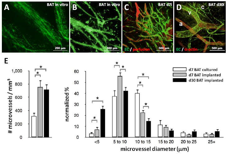
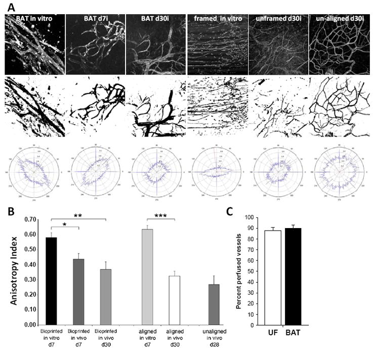
Methods and results: We used 3D direct-write bioprinting or physical constraints in a manner permitting postangiogenesis vascular remodeling and adaptation to pattern angiogenic microvascular precursors (neovessels formed from isolated microvessel segments) in 3D collagen gels before implantation and subsequent network formation. Neovasculatures prepatterned into parallel arrays formed functional networks after 4 weeks postimplantation but lost the prepatterned architecture. However, maintenance of uniaxial physical constraints during postangiogenesis remodeling of the implanted neovasculatures produced networks with aligned microvessels, as well as an altered proportional distribution of arterioles, capillaries, and venules.

Conclusions: Here we show that network topology resulting from implanted microvessel precursors is independent from prepatterning of precursors but can be influenced by a patterning stimulus involving tissue deformation during postangiogenesis remodeling and maturation.


Conflict of interest statement

Competing financial interests: No competing financial interests exist.

Figures

Figure 1
Figure 1
Microvascular constructs formed by 3-D printing using the BAT (BioAssembly Tool) or by physical framing. A) Still image of the final fill stage from a video clip (see Supplementary Movie S1) of MVC printing showing the printing tip (arrow) dispensing collagen:fragments in the hydrogel mold. B) A phase image of a newly bioprinted MVC with microvessel fragments (arrows) dispersed throughout the stripe of collagen. C) Schematic of the different constrained conditions. MVCs constructs were placed in a frame which constrains along the single long axis. Following culture, the construct was either removed from the frame (“unframed”) or kept in the frame (“framed”) for implantation. Un-aligned MVCs were prepared by forming the constructs in well-plates, which constrains radially, and removing them prior to implantation. D) Side view of the stainless steel frame used in the “framed” experiments and made from screen cut in long strips and then folded in at the ends to allow for better construct anchoring. Scale is in centimeters. E) Top view of framed MVCs in culture used for “unframed” implants.
Figure 2
Figure 2
Microvascular networks formed in implanted MVC do not maintain a pre-aligned orientation. Low magnification epifluorescence image (A) and high magnification confocal stack (B) of angiogenic neovessels formed from isolated parent microvessel fragments (isolated from GFP expressing transgenic rats) in BAT-printed MVCs cultured for 7 days. C) Following implantation, the neovessels within the BAT-printed MVC lines have assembled into a simple network and are perfused after 7 days post-implantation (BAT d7i). D) After 30 days post-implantation (BAT d30i), mature arterioles (a), capillaries (c) and venules (v) are present within a hierarchical network. E) The density of microvessels and the % distribution of microvessels of a given diameter in printed MVCs. All values are presented as mean ± standard error of the mean. Statistical significance was determined using one-way ANOVA with post-hoc (p≤ 0.05 was considered significant). Legend applies to both data graphs.
Figure 3
Figure 3
Fast Fourier transform (FFT)-based analysis of microvessel anisotropy in patterned MVCs. A) In the top row are examples of vascular fields from cultured (in vitro) and day 7 (d7i) or day 30 (d30i) implanted MVCs used in the analysis. The middle row contains the corresponding thresholded and binarized images used to generate the orientation plots by FFT (bottom row). B) Plot of anisotropy indices generated from the FFT analysis for each of the conditions. * = p < 0.05, ** = p < 0.01, *** = p < 0.001. C) Percentage of vessels within unframed (UF) and bioprinted (BAT) MVC implants (day 30) perfused with a fluorescent blood tracer (rhodamine-dextran, 3 million MW).
Figure 4
Figure 4
MVC implants with continuous mechanical constraints formed aligned microvascular networks. A) Brightfield image of a framed MVC (dashed outline) explanted after 30 days of subcutaneous implantation. B-D) Stitched confocal projections showing the overall organization of the microvessels in framed cultured MVC (framed in vitro), unframed implants (unframed d30i), and framed implants (framed d30i) prepared from GFP-expressing transgenic rats. E-F) Example FFT plots and anisotropy values for unframed and framed 30 day implants (both framed in culture) showing that continuous framing of the implants maintained microvessel alignment while un-framing did not. Cultured = open bars; implanted = filled bars.
Figure 5
Figure 5
Computer simulations of collagen gel deformation in unframed and framed implants. A) Predicted shape-change and strain maps (top row) and microvessel alignments (bottom row) in cultured MVCs (framed in vitro), unframed and framed implants from computer simulations in which the collagen gel was modeled as a compressible neo-Hookean hyperelastic constitutive system with a viscoelastic component. The color scale for the top panel ranges from blue (negative strain, maximum of -0.3 Pa) to red (positive strain, maximum of +0.43 Pa), green represents zero strain. B) The percentage of neovessels in each simulated condition in 30° angle bins where an alignment of 0° indicates an alignment along the direction of constraint according to the schematic shown to the right. The dashed line indicates a system with randomly oriented elements. Scale bars in panels equal 500 μm. * equals p < 0.05.
Figure 6
Figure 6
Microvessel composition of mechanically loaded MVC implants is different. A) Unframed (A) and framed (B) implants have microvasculatures that are perfused (top) and are comprised of mature arterioles (a), capillaries (c) and venules (v) (bottom). C) Networks in framed MVC implants contained proportionally more capillaries and fewer arterioles than those in unframed MVC implants. D) Microvascular networks of both unframed (UF) and framed (F) implants have equal numbers of microvessels. E-F) Arteriole and venule diameters are significantly larger in the framed networks than in the unframed networks and are perfused to an equal extent. G) Ink casting of the mouse gracilis muscle vasculature highlighting the similarity between the aligned network in skeletal muscle and the aligned network in the framed MVC implants. Scale bars in panels equal 200 μm. * equals p < 0.05.

References

    1. Pittman RN. Oxygen transport and exchange in the microcirculation. Microcirculation. 2005;12:59–70. - PubMed
    1. Pries AR, Secomb TW. Control of blood vessel structure: insights from theoretical models. Am J Physiol Heart Circ Physiol. 2005;288:H1010–H1015. - PubMed
    1. Hoying JB, Williams SK. Building blood vessels. In: Aird WC, editor. Endothelial Biomedicine. Cambridge: Cambridge University Press; 2007.
    1. Nunes SS, Greer KA, Stiening CM, Chen HY, Kidd KR, Schwartz MA, Sullivan CJ, Rekapally H, Hoying JB. Implanted microvessels progress through distinct neovascularization phenotypes. Microvasc Res. 2010;79:10–20. - PMC - PubMed
    1. Pries AR, Cornelissen AJ, Sloot AA, Hinkeldey M, Dreher MR, Hopfner M, Dewhirst MW, Secomb TW. Structural adaptation and heterogeneity of normal and tumor microvascular networks. PLoS Comput Biol. 2009;5:e1000394. - PMC - PubMed

Publication types