. 2012 Feb;40(4):1407-23.
doi: 10.1093/nar/gkr810. Epub 2011 Nov 3.

Comprehensive survey and geometric classification of base triples in RNA structures

Affiliations

Comprehensive survey and geometric classification of base triples in RNA structures

Amal S Abu Almakarem et al. Nucleic Acids Res. 2012 Feb.

Abstract

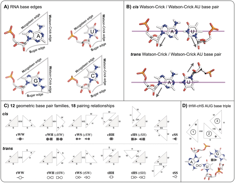
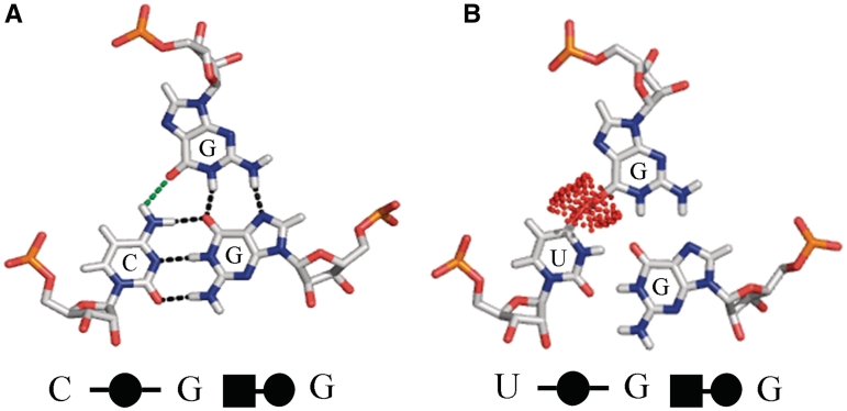
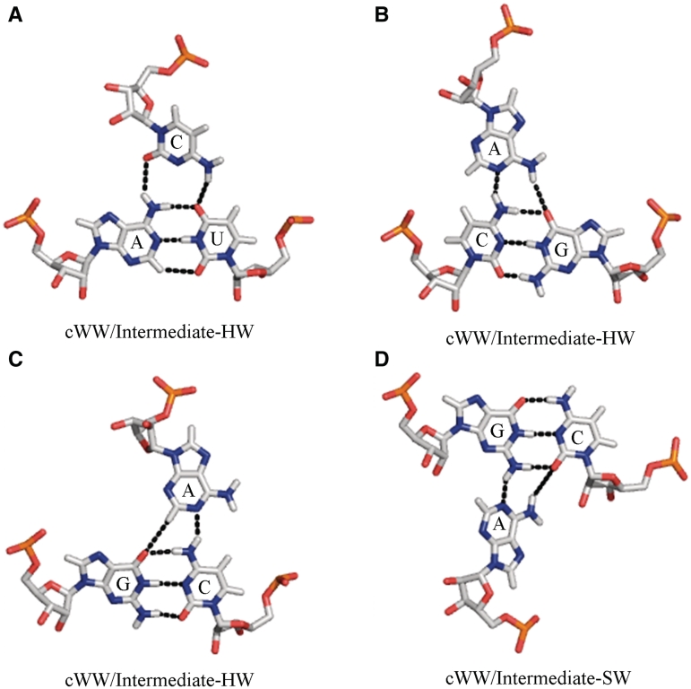
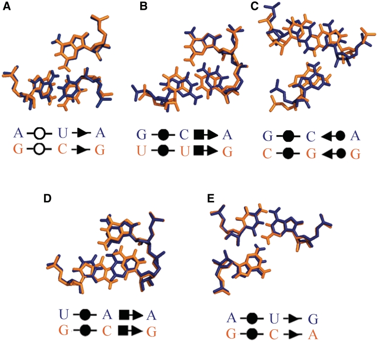
Base triples are recurrent clusters of three RNA nucleobases interacting edge-to-edge by hydrogen bonding. We find that the central base in almost all triples forms base pairs with the other two bases of the triple, providing a natural way to geometrically classify base triples. Given 12 geometric base pair families defined by the Leontis-Westhof nomenclature, combinatoric enumeration predicts 108 potential geometric base triple families. We searched representative atomic-resolution RNA 3D structures and found instances of 68 of the 108 predicted base triple families. Model building suggests that some of the remaining 40 families may be unlikely to form for steric reasons. We developed an on-line resource that provides exemplars of all base triples observed in the structure database and models for unobserved, predicted triples, grouped by triple family, as well as by three-base combination (http://rna.bgsu.edu/Triples). The classification helps to identify recurrent triple motifs that can substitute for each other while conserving RNA 3D structure, with applications in RNA 3D structure prediction and analysis of RNA sequence evolution.


Figures

Figure 1.
Figure 1.
Summary of Leontis/Westhof base pairing classification. (A) Each unmodified RNA nucleotide presents three edges for base pairing interactions, the Hoogsteen (H), Watson–Crick (W) and Sugar (S) edges. Consequently, nucleobases can be conveniently represented by triangles as shown. Note that the sugar edges include the 2′-OH group of the riboses. (B) For each pair of edges, nucleotides can pair in two distinct ways, designated cis and trans, and related by 180° rotation of one nucleotide about the magenta axis that bifurcates the nucleobases perpendicular to the interacting edges. The glycosidic bonds of the nucleotides are on the same side of this axis in the cis configuration, and on opposite sides in the trans configuration (indicated by arrows). (C) Schematic representations of each of the 12 basic base pair families, using triangles to represent each base. Symbols for annotating secondary structures of RNA with non-Watson–Crick base pairs are also provided. The symbols are derived by associating circles with W edges, squares with H edges and triangles with S edges. Filled in symbols represent cis base pairs and open symbols, trans base pairs. Note that the 12 base pair families result in 18 base pairing relations due to the asymmetry of some base pairs. (D) Schematic showing a representative regular base triple, AUG tHW/cHS. The central base (U), numbered base ‘2’, pairs with each of the other two bases of the triple using a distinct base edge. A is base 1 and G is base 3. The triple is named according to the base pairs formed by bases 1 and 2 (tHW in this case) and by bases 2 and 3 (cHS in this case).
Figure 2.
Figure 2.
Examples of favorable (A) and unfavorable (B) interactions between the first and third bases of a triple. (A) The exemplar structure of the cWW/cHW CGG base triple which occurs at higher than expected frequency in the structure database. This triple appears to be stabilized by a favorable interaction between the first and third bases of the triple, the H-bond shown with green dotted line between C(N4) and G(O6) of the third base. (B) The steric clash between the first and third base that prevents the cWW/cHW UGG triple from forming. Red dots define clashing van der Waals surfaces.
Figure 3.
Figure 3.
Examples of intermediate base triples in which bases 1 and 2 are cWW paired and the third base interacts with atoms on the adjoining Hoogsteen or Sugar edges of both bases 1 and 2, without forming individual base pairs with either base. (A) Structure of AUC cWW/Intermediate-HW triple A2882/U2836/C2879 from 23S rRNA, PDB file 3I8I (27). (B) Structure of CGA cWW/Intermediate-HW triple G22/C43/A9 from PDB file 2QWY (40). (C) Structure of GCA cWW/Intermediate-HW triple G2580/C2555/A2577 from PDB file 1S72 (42). (D) Structure of GCA cWW/ Intermediate-SW triple G769/C810/A900 from 1J5E (26). In panels (A), (B), and (C) the Watson-Crick edge of the third base interacts with atoms on the Hoogsteen edges of bases 1 and 2, without forming individual base pairs with either one. In panel (D) the Watson-Crick edge of the third base interacts with atoms on the Sugar edges of bases 1 and 2, again without forming individual base pairs with either one.
Figure 4.
Figure 4.
Examples of covariation of conserved base triples observed at corresponding positions in E. coli (blue) and T. thermophilus (orange) from 23S rRNA (Upper row) and 16S rRNA (Lower row). Upper row (A): Conserved tWW/cSS base triple in 23S rRNA nucleotides 430/234/219, which is AUA in E.c. and GCG in T. th. (B): Conserved cWW/cHS base triple in 23S rRNA position 757/740/739, which is GCA in E.c. and UUG in T. th. (C): Conserved cWW/cSW base triple in 23S rRNA position 418/409/226, which is GCA in E.c. and CGG in T. th. Lower row (D): Conserved cWW/cHS base triple in 16S rRNA position 644/595/596, which is UAA in E.c. and GCG in T. th. (E): Conserved cWW/cSS base triple in 16S rRNA position 1326/1311/1268, which is AUG in E. coli and GCA in T. th. Structures were taken from the following PDB files: E. coli 23S rRNA, 2QBE (25); E. coli 16S rRNA, 2QAN (25) T. th. 23S rRNA, 3I8I (27) and T. th. 16S rRNA, 1J5E (26).

References

    1. Leontis NB, Westhof E. A common motif organizes the structure of multi-helix loops in 16 S and 23 S ribosomal RNAs. J. Mol. Biol. 1998;283:571–583. - PubMed
    1. Szewczak AA, Moore PB. The sarcin/ricin loop, a modular RNA. J. Mol. Biol. 1995;247:81–98. - PubMed
    1. Klein DJ, Schmeing TM, Moore PB, Steitz TA. The kink-turn: a new RNA secondary structure motif. Embo J. 2001;20:4214–4221. - PMC - PubMed
    1. Lescoute A, Leontis NB, Massire C, Westhof E. Recurrent structural RNA motifs, Isostericity Matrices and sequence alignments. Nucleic Acids Res. 2005;33:2395–2409. - PMC - PubMed
    1. Doherty EA, Batey RT, Masquida B, Doudna JA. A universal mode of helix packing in RNA. Nat. Struct. Biol. 2001;8:339–343. - PubMed

Publication types