

Cerebral actinomycosis : unusual clinical and radiological findings of an abscess
- PMID: 22053238
- PMCID: PMC3206280
- DOI: 10.3340/jkns.2011.50.2.147
Cerebral actinomycosis : unusual clinical and radiological findings of an abscess
Abstract
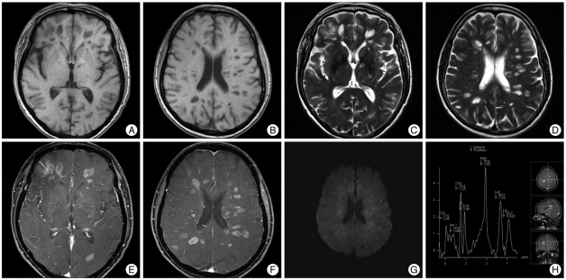
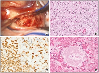
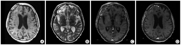
We report a case of cerebral actinomycosis in a 69-year-old immunocompetent woman. The patient showed a progressive worsened mental status for one week. MRI examination showed an increased size of multiple enhancing nodular lesions associated with mild perilesional edema. We performed an open biopsy for the right frontal enhancing lesion. The intraoperative finding showed a yellowish friable lesion that was not demarcated with normal tissue. Pathologically, an actinomycotic lesion with sulfur granules and inflammatory cells was diagnosed. We report an unusual case of diffuse involvement of cerebral actinomycosis. The presence of the uncapsulated friable lesion that consisted mainly of foamy macrophages and lymphocytes could explain the unusual radiological features.
Keywords: Actinomycosis; Cerebral abscess; MR imaging.
Figures
References
-
- Adeyemi OA, Gottardi-Littell N, Muro K, Kane K, Flaherty JP. Multiple brain abscesses due to Actinomyces species. Clin Neurol Neurosurg. 2008;110:847–849. - PubMed
-
- Alday R, Lopez-Ferro MO, Fernandez-Guerrero M, Ruiz-Barnés P. Spinal intrarhecal empyema due to Actinomyces israeli. Acta Neurochir (Wien) 1989;101:159–162. - PubMed
-
- Al-Okaili RN, Krejza J, Wang S, Woo JH, Melhern ER. Advanced MR imaging techniques in the diagnosis of intraaxial brain tumors in adults. Radiographics. 2006;26(Suppl 1):S173–S189. - PubMed
-
- Bébrová E, Lochmann O, Tichý M, Nyc O. [Actinomyces viscosus in subdural empyema] Cesk Epidemiol Mikrobiol Imunol. 1994;43:21–22. - PubMed
-
- Brown JR. Human actinomycosis. A study of 181 subjects. Hum Pathol. 1973;4:319–330. - PubMed
Publication types
LinkOut - more resources
Full Text Sources
