. 2011 Nov 4:4:20.
doi: 10.1186/1756-8935-4-20.

Myc and Miz-1 have coordinate genomic functions including targeting Hox genes in human embryonic stem cells

Affiliations

Myc and Miz-1 have coordinate genomic functions including targeting Hox genes in human embryonic stem cells

Natalia Varlakhanova et al. Epigenetics Chromatin. .

Abstract

Background: A proposed role for Myc in maintaining mouse embryonic stem (ES) cell pluripotency is transcriptional repression of key differentiation-promoting genes, but detail of the mechanism has remained an important open topic.

Results: To test the hypothesis that the zinc finger protein Miz-1 plays a central role, in the present work we conducted chromatin immunoprecipitation/microarray (ChIP-chip) analysis of Myc and Miz-1 in human ES cells, finding homeobox (Hox) genes as the most significant functional class of Miz-1 direct targets. Miz-1 differentiation-associated target genes specifically lack acetylated lysine 9 and trimethylated lysine 4 of histone H3 (AcH3K9 and H3K4me3) 9 histone marks, consistent with a repressed transcriptional state. Almost 30% of Miz-1 targets are also bound by Myc and these cobound genes are mostly factors that promote differentiation including Hox genes. Knockdown of Myc increased expression of differentiation genes directly bound by Myc and Miz-1, while a subset of the same genes is downregulated by Miz-1 loss-of-function. Myc and Miz-1 proteins interact with each other and associate with several corepressor factors in ES cells, suggesting a mechanism of repression of differentiation genes.

Conclusions: Taken together our data indicate that Miz-1 and Myc maintain human ES cell pluripotency by coordinately suppressing differentiation genes, particularly Hox genes. These data also support a new model of how Myc and Miz-1 function on chromatin.


Figures
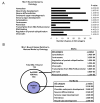
Figure 1
Figure 1
Genomic binding of Miz-1 and functional ontology of its target genes in human embryonic stem (ES) cells. (A) Gene ontology (GO) functional annotation of Miz-1 bound genes was performed using DAVID functional annotation software. The most significant biological process GO terms are shown. The number of genes belonging to each category within the Miz-1 bound population is indicated by black bars. (B) Acetylated lysine 9 and trimethylated lysine 4 of histone H3 (AcH3K9 and H3K4me3) status for Miz-1 bound target genes. P values are indicated in all cases.
Figure 2
Figure 2
Genomic binding of Myc and functional ontology of its target genes in human embryonic stem (ES) cells. (A) Acetylated lysine 9 and trimethylated lysine 4 of histone H3 (AcH3K9 and H3K4me3) status for Myc bound genes. Gene ontology analysis for Myc bound genes with or without enrichment in histone activation marks H3K4me3 and AcH3K9 was performed using DAVID functional annotation software. P values are indicated. Myc binding events that do not overlap with either histone modification are represented by the blue circle. (B) Confirmation of Myc direct targets identified by chromatin immunoprecipitation/microarray (ChIP-chip). ChIP was performed using antibodies to c-Myc, N-Myc, AcH3K9, and H3K4me3. PR2Y2 gene, not bound by either c-Myc or N-Myc, was used as a negative control.
Figure 3
Figure 3
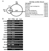
Myc and Miz-1 are cobound on differentiation-associated genes. (A) Venn diagram showing overlap of Miz-1 and Myc bound genes. The most significant biological process gene ontology (GO) terms are shown on the right with P values. (B) Chromatin immunoprecipitation (ChIP) confirmation of Miz-1 and Myc common target genes identified by ChIP/microarray (ChIP-chip) analysis. SnurpN and OSRF genes, identified to be bound respectively only by Miz-1 or only by N-Myc by ChIP-chip, were used as a negative control.
Figure 4
Figure 4
Target genes bound by both Myc and Miz-1 have a distinct, transcriptionally inactive profile of histone marks. (A) Examples of enrichment profiles observed for Myc, Miz-1, acetylated lysine 9 of histone H3 (AcH3K9) (AcK9) and trimethylated lysine 4 of histone H3 (H3K4me3) (TMK4) for Myc/Miz-1, Miz-1 alone or Myc alone bound genes are shown. Miz-1, Myc, TMK4, and AcK9 are indicated in blue, red, green, and purple, respectively. Black horizontal arrows indicate direction of transcription. Peaks are on a Log2 scale. Gene names are indicated above the colored horizontal bars with tiled regions immediately below and transcriptional start sites (TSSs) are indicated by short vertical lines near the bottom. (B) Chromatin immunoprecipitation (ChIP) analysis of H3K27me3 enrichment on Myc/Miz-1 and Myc alone bound genes. (C) Myc and Miz-1 binding distribution relative to the TSS.
Figure 5
Figure 5
Loss of Myc leads to spontaneous differentiation and upregulation of differentiation-associated genes. (A) Confirmation of c-Myc or N-Myc knockdown by quantitative real-time (qRT)-PCR PCR. Levels of Myc RNAs measured by qRT-PCR were normalized to the levels of loading control peptidylprolyl isomerase A (PPIA; cyclophilin A). Error bars are standard deviations. N = 3. (B) Alkaline phosphatase staining of vector alone or Myc small hairpin (sh)RNA human embryonic stem (ES) cell colonies. Images were taken at either 4 × (top panels) or 10 × magnifications (bottom panels). (C) Gene ontology (GO) analysis of genes that are differentially expressed upon Myc knockdown with indicated P values.
Figure 6
Figure 6
Myc represses expression of differentiation-associated including homeobox (Hox) genes, a function antagonized by Miz-1. (A) Confirmation of gene expression changes in c-myc knockdown (KD) embryonic stem (ES) cells, determined by gene expression array, by quantitative real-time (qRT)-PCR PCR. Real-time qRT-PCR of a series of differentiation-associated genes or metabolism related genes. Error bars are standard deviations. N = 3. (B) Venn diagram showing overlap among differentially expressed genes in Myc KD or Miz-1 KD ES cells. Gene ontology (GO) analysis of overlapping genes presented below the Venn diagram. P values are indicated.
Figure 7
Figure 7
Model. Myc/Miz-1 pathway acts as a master switch that represses multiple differentiation-associated genes.

References

    1. Peukert K, Staller P, Schneider A, Carmichael G, Hanel F, Eilers M. An alternative pathway for gene regulation by Myc. EMBO J. 1997;16:5672–5686. doi: 10.1093/emboj/16.18.5672. - DOI - PMC - PubMed
    1. Gartel AL, Shchors K. Mechanisms of c-myc-mediated transcriptional repression of growth arrest genes. Exp Cell Res. 2003;283:17–21. doi: 10.1016/S0014-4827(02)00020-4. - DOI - PubMed
    1. Staller P, Peukert K, Kiermaier A, Seoane J, Lukas J, Karsunky H, Möröy T, Bartek J, Massague J, Hänel F, Eilers M. Repression of p15INK4b expression by Myc through association with Miz-1. Nat Cell Biol. 2001;3:392–399. doi: 10.1038/35070076. - DOI - PubMed
    1. Wanzel M, Russ AC, Kleine-Kohlbrecher D, Colombo E, Pelicci P-G, Eilers M. A ribosomal protein L23-nucleophosmin circuit coordinates Miz1 function with cell growth. Nat Cell Biol. 2008;10:1051–1061. doi: 10.1038/ncb1764. - DOI - PubMed
    1. Seoane J, Le HV, Massague J. Myc suppression of the p21(Cip1) Cdk inhibitor influences the outcome of the p53 response to DNA damage. Nature. 2002;419:729–734. doi: 10.1038/nature01119. - DOI - PubMed