. 2012 Jan;83(1):41-51.
doi: 10.1111/j.1365-2958.2011.07909.x. Epub 2011 Nov 22.

Surface contact stimulates the just-in-time deployment of bacterial adhesins

Affiliations

Surface contact stimulates the just-in-time deployment of bacterial adhesins

Guanglai Li et al. Mol Microbiol. 2012 Jan.

Abstract

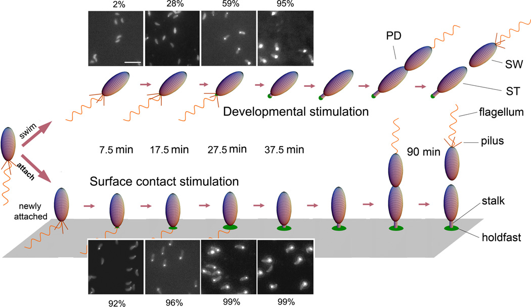
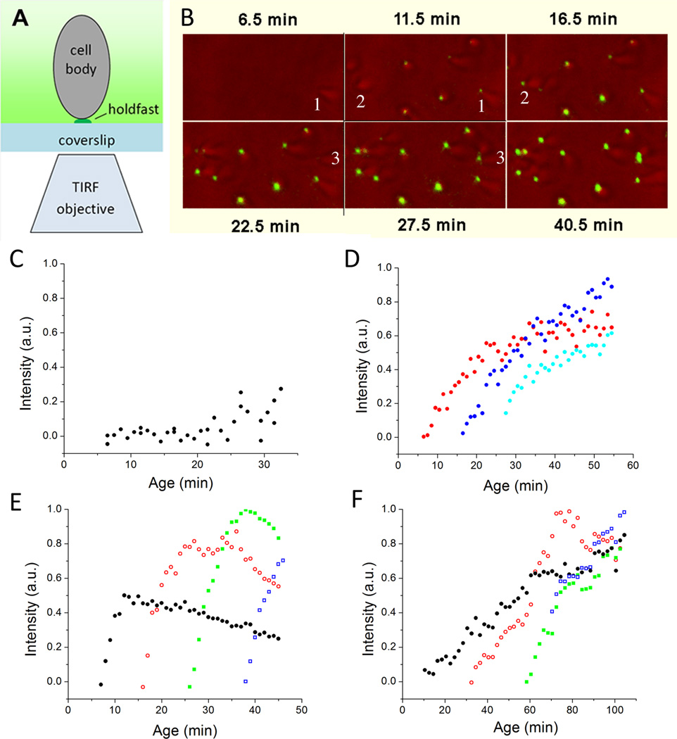
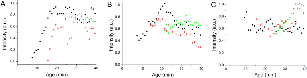
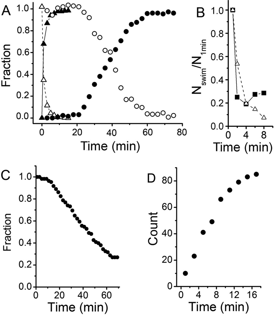
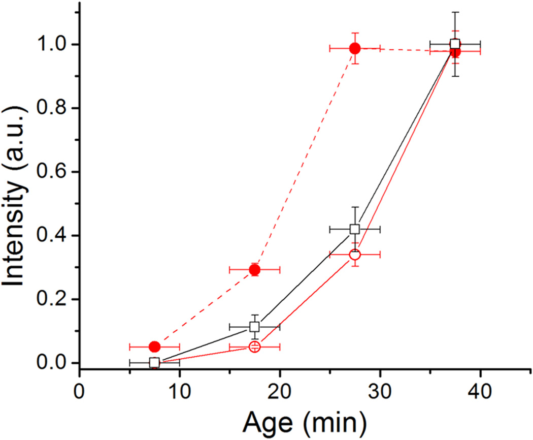
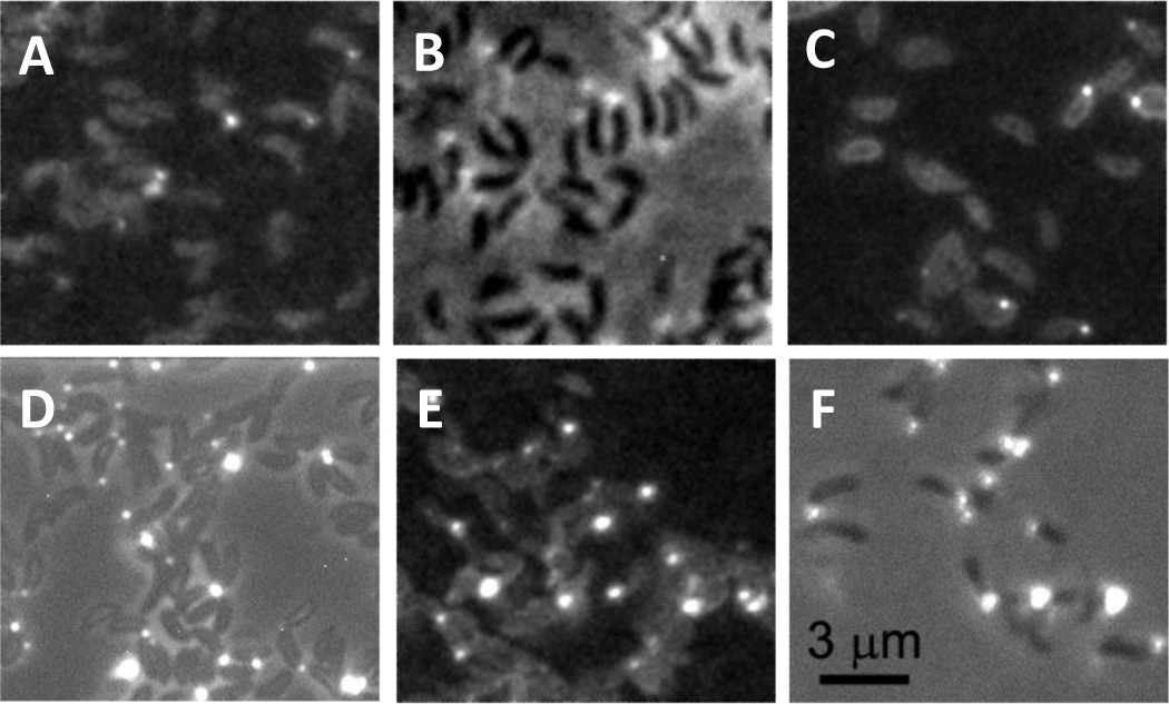
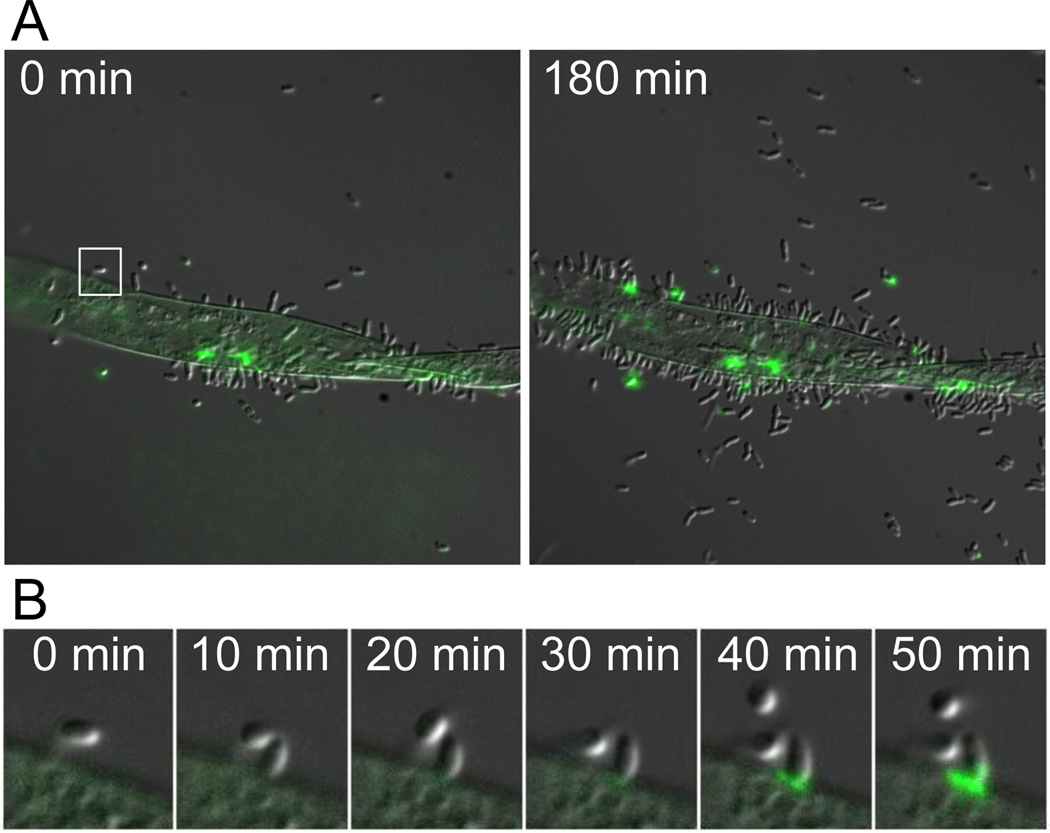
The attachment of bacteria to surfaces provides advantages such as increasing nutrient access and resistance to environmental stress. Attachment begins with a reversible phase, often mediated by surface structures such as flagella and pili, followed by a transition to irreversible attachment, typically mediated by polysaccharides. Here we show that the interplay between pili and flagellum rotation stimulates the rapid transition between reversible and polysaccharide-mediated irreversible attachment. We found that reversible attachment of Caulobacter crescentus cells is mediated by motile cells bearing pili and that their contact with a surface results in the rapid pili-dependent arrest of flagellum rotation and concurrent stimulation of polar holdfast adhesive polysaccharide. Similar stimulation of polar adhesin production by surface contact occurs in Asticcacaulis biprosthecum and Agrobacterium tumefaciens. Therefore, single bacterial cells respond to their initial contact with surfaces by triggering just-in-time adhesin production. This mechanism restricts stable attachment to intimate surface interactions, thereby maximizing surface attachment, discouraging non-productive self-adherence, and preventing curing of the adhesive.


Figures

Figure 1
Figure 1
Cell cycle diagram and holdfast production of unattached or attached C. crescentus cells. Fluorescence images show unattached (top) and attached (bottom) synchronized C. crescentus wild-type cells labeled with fluorescein-WGA and imaged at ages 7.5 ± 2.5 min, 17.5 ± 2.5 min, 27.5 ± 2.5 min, and 37.5 ± 2.5 min. The contrast of the images was scaled up to show the dim cell body due to non-specific labeling. The bright spot at the pole of a cell body is the holdfast. The percentage of cells with a holdfast at each time point is indicated. Attached cells rapidly produce a holdfast after surface contact, whereas unattached swarmer cells produce a holdfast just prior to differentiating into stalked cells.
Figure 2
Figure 2
Holdfast production observed for individual cells with TIRF microscopy. A) Schematic drawing showing the configuration of the TIRF microscopy and a cell attached to the coverslip. Only the fluor (shown in green) within a couple hundred nanometers from the coverslip is visible. B) Overlay of light and TIRF images obtained during a time course with wild-type C. crescentus. Cells arrive at the surface without a holdfast but the holdfast is detected in the next image, which was taken five min later (see numbered examples). C) Integrated intensity in arbitrary unit (a.u.) of the TIRF image of the holdfast of newly attached wild-type C. crescentus cells. Each black dot represents the data from a newly attached individual cell. D) Integrated intensity of the TIRF images of the holdfasts of three wild-type C. crescentus CB15 cells after attachment at an age of 6.5 min (red), 16.5 min (blue), and 26.5 min (cyan). Each color shows the fluorescence intensity for a single cell as it varies through time. E) Integrated intensity of the TIRF images of the holdfasts of four A. biprosthecum cells after attachment at an age of 7.5 min (black), 16.5 min (red), 26.5 min (green), and 38.5 min (blue). Each color shows the fluorescence intensity for a single cell as it varies through time. The fluorescence intensity eventually declines due to photobleaching. F) Integrated intensity of TIRF image of the holdfasts of four A. tumefaciens cells after attachment at an age of 10.5 min (black), 32.5 min (red), 58.5 min (green), and 70.5 min (blue). Each color shows the fluorescence intensity for a single cell as it varies through time.
Figure 3
Figure 3
Integrated intensity of the TIRF images of the holdfasts of A) three C. crescentus wild-type cells in medium with 10 µg/ml kanamycin at ages of 7.5 min (black), 15.5 min (red) and 26.5 min (green); B) three C. crescentus wild-type cells in medium with 10% (w/v) Ficoll 400 attached at an age of 7.5 min (black), 14.5 min (red), and 21.5 min (green); C) three C. crescentus ΔpilA cells in medium with 10% (w/v) Ficoll 400 attached at an age of 9.5 min (black), 15.5 min (red), and 25.5 min (green).
Figure 4
Figure 4
Attachment of C. crescentus strains CB15 (wild-type), ΔpilA, and ΔhfsA to glass surfaces. A) The fraction of attached (solid symbol) and swimming cells (open symbol) in the field of view at various times after the synchronized cells were placed between two glass surfaces separated by 12 µm. Synchronized wild-type cells (triangle) attached at a rate of 68% of cells per min immediately after birth and almost all of them had attached to the glass surface within 5 min. Synchronized ΔpilA cells (circle) only began to attach after 20 min and their attachment occurred at a rate of 2.8% of cells per min. B) Attachment of C. crescentus wild-type (triangle) and ΔhfsA (square) cells. Since the ΔhfsA strain cannot be synchronized the number of swimming cells in mixed populations (containing swarmer, stalked and predivisional cells) of both strains was normalized to that at 1 min as a function of time after the culture at mid-exponential phase was placed between two glass surfaces separated by 12 µm. C) Fraction of C. crescentus ΔhfsA cells attached to a surface decreases over time as observed under light microscopy. In a similar experiment, wild-type cells remained attached to the surface for at least 90 min (data not shown). D) Number of cells from mixed culture of a ΔpilA ΔflgE double mutant attaching to a surface. Cells were grown to mid-exponential phase, placed between two coverslips, and a 40 X objective was focused on the bottom surface to take videos. Because these cells do not swim, most cells settled on the bottom surface. Unattached cells were distinguished from attached cells because unattached cells are subject to Brownian motion while attached cells are not.
Figure 5
Figure 5
Integrated fluorescence intensity of holdfast of unattached wild-type cells (open circle), wild-type attached cells (solid circle), and unattached ΔpilA cells (open square) at different ages. The error bar is the standard error of 2 to 4 measurement for each data point. Age refers to the time after cell division.
Figure 6
Figure 6
Holdfast of cells at age 17.5 min grown in A) PYE medium, B) PYE medium with 67% Percoll, C) PYE medium with 3.5% PEG 8000, D) PYE medium with 5% PEG 35000, E) PYE medium with 5% Dextran, and F) PYE medium with 10% Ficoll. Panels A, C, and E are fluorescence images, which clearly show the cell bodies in addition to the holdfasts. Panels B, D, and F are overlays of fluorescence and phase contrast images.
Figure 7
Figure 7
Timelapse microscopy of Agrobacterium tumefaciens cells attaching to Arabidopsis roots. Production of UPP was monitored using WGA-AlexaFluor 488. A) At the initial time point, few cells are attached to the root and little UPP is detected. After 180 min, many more cells are attached to the root and UPP is frequently detected along the junction between the bacterial cells and the root. UPP is rarely observed in the unattached cells. B) Close-up of the region boxed in A. A single A. tumefaciens cell attaches to the root and subsequent production of UPP is observed.

Comment in

References

    1. Agladze K, Wang X, Romeo T. Spatial periodicity of Escherichia coli K-12 biofilm microstructure initiates during a reversible, polar attachment phase of development and requires the polysaccharide adhesin PGA. J Bacteriol. 2005;187:8237–8246. - PMC - PubMed
    1. Anderson JK, Smith TG, Hoover TR. Sense and sensibility: flagellum-mediated gene regulation. Trends Microbiol. 2010;18:30–37. - PMC - PubMed
    1. Beloin C, Houry A, Froment M, Ghigo JM, Henry N. A short-time scale colloidal system reveals early bacterial adhesion dynamics. PLoS Biol. 2008;6:e167. - PMC - PubMed
    1. Blair KM, Turner L, Winkelman JT, Berg HC, Kearns DB. A molecular clutch disables flagella in the Bacillus subtilis biofilm. Science. 2008;320:1636–1638. - PubMed
    1. Bodenmiller D, Toh E, Brun YV. Development of surface adhesion in Caulobacter crescentus. J.Bacteriol. 2004;186:1438–1447. - PMC - PubMed

Publication types

MeSH terms

Substances