Review
. 2011 Nov;3(11):1317-40.
doi: 10.2217/imt.11.115.

Chemokines, costimulatory molecules and fusion proteins for the immunotherapy of solid tumors

Affiliations
Review

Chemokines, costimulatory molecules and fusion proteins for the immunotherapy of solid tumors

Melissa G Lechner et al. Immunotherapy. 2011 Nov.

Abstract

In this article, the role of chemokines and costimulatory molecules in the immunotherapy of experimental murine solid tumors and immunotherapy used in ongoing clinical trials are presented. Chemokine networks regulate physiologic cell migration that may be disrupted to inhibit antitumor immune responses or co-opted to promote tumor growth and metastasis in cancer. Recent studies highlight the potential use of chemokines in cancer immunotherapy to improve innate and adaptive cell interactions and to recruit immune effector cells into the tumor microenvironment. Another critical component of antitumor immune responses is antigen priming and activation of effector cells. Reciprocal expression and binding of costimulatory molecules and their ligands by antigen-presenting cells and naive lymphocytes ensures robust expansion, activity and survival of tumor-specific effector cells in vivo. Immunotherapy approaches using agonist antibodies or fusion proteins of immunomodulatory molecules significantly inhibit tumor growth and boost cell-mediated immunity. To localize immune stimulation to the tumor site, a series of fusion proteins consisting of a tumor-targeting monoclonal antibody directed against tumor necrosis and chemokines or costimulatory molecules were generated and tested in tumor-bearing mice. While several of these reagents were initially shown to have therapeutic value, combination therapies with methods to delete suppressor cells had the greatest effect on tumor growth. In conclusion, a key conclusion that has emerged from these studies is that successful immunotherapy will require both advanced methods of immunostimulation and the removal of immunosuppression in the host.


Figures

Figure 1
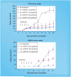
Figure 1. CCL16 immunotherapy increases tumor infiltration by immune cells and inhibits growth of established tumors
(A) CCL16 treatment of CT26 tumors produced significant influx of immune cells into the tumor site, most notably DCs and granulocytes (original magnification ×200). (B) Both targeted (LEC-chTNT-3) and untargeted (LEC-Fc) CCL16 fusion proteins significantly inhibited CT26 tumor growth in BALB/c mice. For these studies, tumor-bearing mice (n = 5) were injected intravenously with 25 μg of LEC reagents daily for 5 days starting on day 6 after tumor implantation. DC: Dendritic cell; E: Eosin; H: Hematoxylin; LEC: Liver-expressed chemokine; PMN: Polymorphonuclear cell; TNT: Tumor necrosis therapy.
Figure 2
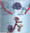
Figure 2. Chemokines for cancer immunotherapy and their role in cancer metastasis
(A) Chemokine immunotherapy to promote naive and activated immune-cell infiltration into the tumor microenvironment. One challenge of using chemokines for immunotherapy is their dual actions in recruiting immune effector and immune suppressor cells. Chemokine immunotherapy may be paired with strategies to boost tumor antigen capture and presentation for robust antitumor immunity. (B) Aberrant overexpression of chemokine receptors mediates tumor cell migration and seeding of metastatic disease. CCL19 and CCL21 facilitate homing of naive lymphocytes and mature APCs to LNs under normal conditions, but can be used by CCR7+ tumor cells to migrate to tumor draining LNs. CXCL12 normally regulates immune cell homeostasis in and out of the bone marrow and is co-opted by CXCR4+/CXCR7+ tumor cells for metastasis to bone. APC: Antigen presenting cell; DC: Dendritic cell; iDC: Immature dendritic cell; LN: Lymph node; Mϕ: Macrophage; MDSC: Myeloid-derived suppressor cell; TAA: Tumor-associated antigen; Th: T helper cell; TLR: Toll-like receptor.
Figure 3
Figure 3. Dosing studies of B7.1-Fc and B7.2-Fc immunotherapy in the CT26 tumor model
In these in vivo studies, both (A) B7.1-Fc and (B) B7.2-Fc showed a dose response effect to inhibit tumor growth in this murine solid tumor model. All mice (n = 5) received five daily intravenous treatments starting on day 6 after tumor implantation. Ab: Antibody.
Figure 4
Figure 4. Dendritic cell-T cell synapse
During T-cell docking to dendritic cells, different costimulatory molecules migrate into the synapse to stabilize the cell-cell interaction required for T-cell activation to the peptide presented in the MHC cleft. Without this stabilization, the synapse is too unstable and the cells undock, producing T-cell anergy. CTLA-4 is the inhibitory molecule that, if present, preferentially binds B7.1, thereby preventing T-cell activation.
Figure 5
Figure 5. Fc-mOX40L immunotherapy of CT26 and RENCA tumors in mice
These studies demonstrate that both tumor models (A & B) do not have OX40L costimulation that, when provided, facilitates an effective tumor response by the immune system. OX86, an agonist antibody to OX40, was found to be less effective than the ligand reagents by these methods. All mice (n = 5) received five daily intravenous treatments starting on day 6 after tumor implantation.
Figure 6
Figure 6. Fc-mGITRL immunotherapy of CT26 and RENCA murine tumor models
Dosing studies of Fc-mGITRL showed a dose response in both tumor models. Based upon these results, the CT26 tumor model (A) was found to be more sensitive to Fc-mGITRL treatment than the RENCA tumor model (B) in BALB/c mice. All mice (n = 5) received five daily intravenous treatments starting on day 6 after tumor implantation.
Figure 7
Figure 7. Tumor-targeted chemokines and costimulatory ligands using tumor necrosis treatment monoclonal antibody fusion proteins
Depending upon the location of the active site of the chemokine or costimulatory molecule, each were genetically linked to either the C- or N-terminus of the heavy chain of TNT to produce active fusion proteins. Fc-linked reagents show no specificity for tumor as demonstrated by biodistribution studies and therefore had a more systemic effect when administered intravenously than TNT fusion proteins. LEC: Liver-expressed chemokine; TNT: Tumor necrosis therapy.
Figure 8
Figure 8. Tumor-targeted mCD137L fusion protein immunotherapy shows greater antitumor efficacy compared with untargeted Fc-mCD137L in CT26 tumor-bearing mice
In these studies, groups of tumor-bearing mice (n = 5) received five daily doses of reagent intravenously, starting on day 5 after tumor implantation. Unlike the OX40L results shown in Figure 5, the results indicated that the agonist antibody 2A was the most effective in inhibiting tumor growth in these studies. However, mice receiving chTNT-3/mCD137L showed better tumor inhibition than untargeted Fc-mCD137L, suggesting that that targeting this costimulatory molecule to the tumor microenvironment is more effective for treatment. PBS: Phosphate buffered saline.
Figure 9
Figure 9. Effect of combination B7.1-Fc immunotherapy and CD25+ Treg depletion on the growth of RENCA and MAD109 murine solid tumors (n = 5)
These studies (A & B) demonstrate that combination therapy to eliminate Tregs and provide missing costimulation with B7.1-Fc produces a more profound inhibition of tumor growth in two murine solid tumor models. Treg depletion was accomplished by administering a single 500-μg ip. dose of the rat anti-CD25 monoclonal antibody PC61 on day 0. Ab: Antibody.
Figure 10
Figure 10. Complete regression of established CT26 tumors by combination LEC-chTNT-3 and Treg depletion immunotherapy
Depletion studies with cytotoxic rat antimouse lymphoid antisera demonstrated that removal of CD8+ and NK cells prior to LEC immunotherapy prevented active tumor inhibition. By contrast, depletion of either (A) CD4+ T helper/suppressors cells or (B) CD25+ Tregs produced complete tumor regression when used in combination with LEC-chTNT-3 immunotherapy. In these experiments, the depleting rat antimouse antibodies were administered ip. as a single 500-μg dose on day 0. Groups of mice (n = 5) were then measured for tumor growth by caliper measurement three times per week. As shown in (C), combination immunotherapy with tumor-targeted chemokine CCL16 (LEC-chTNT-3) and anti-CD4+ or anti-CD25+ monoclonal antibodies for Treg cell depletion produced complete regression of established tumors in all experimental animals. In comparison, monotherapy with either LEC-chTNT-3 or Treg cell depletion yielded significant reduction in tumor growth relative to untreated animals but no complete tumor cures. LEC: Liver-expressed chemokine; TNT: Tumor necrosis therapy.
Figure 11
Figure 11. Optimal cancer immunotherapy combines targeted immune stimulation and reversal of tumor immune tolerance
Tumor immune surveillance mechanisms, including antigen-presenting cells (DCs, Mϕ and effector cells [Th cells, CTLs and NK cells]), recognize and attack neoplastic cells in the normal host to control tumor growth. When tumor immune escape occurs, immune suppressor cells recruited to the tumor microenvironment and secondary lymphoid tissues inhibit cell-mediated immunity to facilitate tumor progression. Immune suppressor cells that accumulate in the tumor setting include derivatives of innate (MDSCs, plasmacytoid DCs and tumor-associated Mϕ) and adaptive, or induced, Tregs. In addition to the naturally occurring Treg populations that inhibit reactions to self-antigens at steady state, induced Tregs arise in the periphery in the setting of chronic inflammation and cancer. Finally, there is a major shift in immune cytokines from cell-mediated promoting (Th1) cytokines (IFN-γ, TNF-α and IL-2) to humoral immunity promoting (Th2) cytokines (IL-4, IL-5 and IL-10) in the cancer setting. Immune stimulatory treatments, such as chemokines and costimulatory molecule agonists or ligands, increase tumor infiltrating leukocytes and antigen priming of effector cells. Spatial targeting of immunotherapy to the tumor microenvironment and specificity for tumor-associated antigens can decrease the risk of autoimmune reactions, increase retention time in the tumor and improve the efficacy of treatment. CTL: Cytotoxic T lymphocyte; DC: Dendritic cell; Mϕ: Macrophage; MDSC: Myeloid-derived suppressor cell; Th: T helper cell.

References

    1. Lizée G, Cantu MA, Hwu P. Less yin, more yang: confronting the barriers to cancer immunotherapy. Clin. Cancer Res. 2007;13(18 Pt 1):5250–5255. - PubMed
    1. Stewart TJ, Abrams SI. How tumours escape mass destruction. Oncogene. 2008;27:5894–5903. ■ Excellent primer on tumor immune escape for clinicians and basic scientists, written by an expert in the field of immunotherapy.

    1. Stewart TJ, Smyth MJ. Improving cancer immunotherapy by targeting tumor-induced immune suppression. Cancer Metast. Rev. 2011;30(1):125–140. - PubMed
    1. Sadun RE, Sachsman SM, Chen X, et al. Immune signatures of murine and human cancers reveal unique mechanisms of tumor escape and new targets for cancer immunotherapy. Clin. Cancer Res. 2007;13:4016–4025. ■ Ground-breaking publication showing how identification of the major mechanisms of immune escape in colorectal and breast cancer can be used to tailor successful immunotherapy approaches.

    1. Goldman B, DeFrancesco L. The cancer vaccine roller coaster. Nat. Biotechnol. 2009;27(2):129–139. - PubMed