

Laparoscopic myomectomy of a subserous pedunculated fibroid at 14 weeks of pregnancy: a case report
- PMID: 22054171
- PMCID: PMC3225401
- DOI: 10.1186/1752-1947-5-545
Laparoscopic myomectomy of a subserous pedunculated fibroid at 14 weeks of pregnancy: a case report
Abstract
Introduction: Uterine leiomyomas are seen in 1.6% to 4% of pregnancies. With the increasing age of obstetric patients, more cases are being encountered during pregnancy.
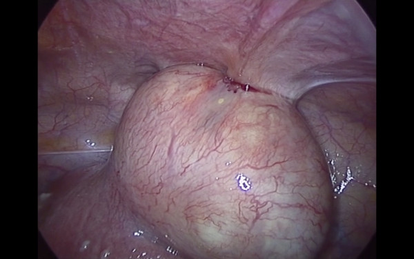
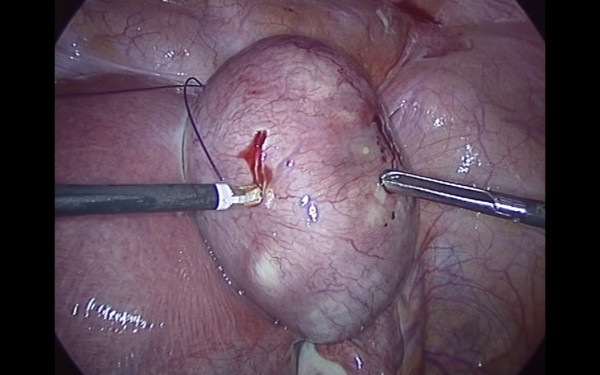
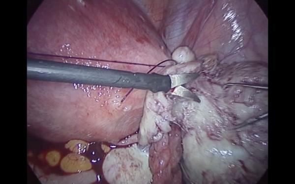
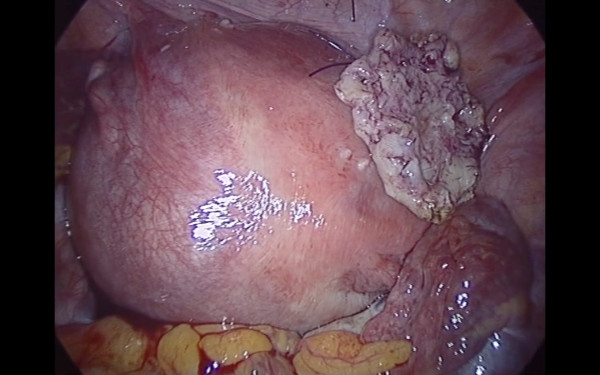
Case presentation: We report the case of a 31-year-old Caucasian woman with acute recurrent abdominal pain due to a subserous fundic myoma, measuring 48 × 52 × 63 mm, with an implantation base of 22 × 18 mm, which was successfully treated by laparoscopy at 14 weeks of pregnancy. At a gestational age of week 40, the patient spontaneously delivered a healthy 3216 g girl baby.
Conclusion: As far as we know, this is the first reported case of laparoscopic myomectomy this early during a pregnancy. Our experience together with an analysis of cases reported in the literature suggests that myomectomy during pregnancy may be considered safe, but only in the hands of experienced laparoscopic surgeons. There are a few reports in the literature about laparoscopic myomectomy during the first half of pregnancy that demonstrate its feasibility in selected cases. Some technical tools could improve the procedure with a minimum of risk for the ongoing pregnancy.
Figures
References
-
- Guidelines Committee of the Society of American Gastrointestinal and Endoscopic Surgeons. Yumi H. Guidelines for diagnosis, treatment, and use of laparoscopy for surgical problems during pregnancy. Surg Endosc. 2008;22:849–861. - PubMed
-
- Bonito M, Gulemì L, Basili R, Roselli D. Myomectomy during the first and second trimester of pregnancy. Clin Exp Obstet Gynecol. 2007;34:149–150. - PubMed
LinkOut - more resources
Full Text Sources
Research Materials
