Review
. 2012:74:299-323.
doi: 10.1146/annurev-physiol-020911-153238. Epub 2011 Nov 4.

Cell biology and pathology of podocytes

Affiliations
Review

Cell biology and pathology of podocytes

Anna Greka et al. Annu Rev Physiol. 2012.

Abstract

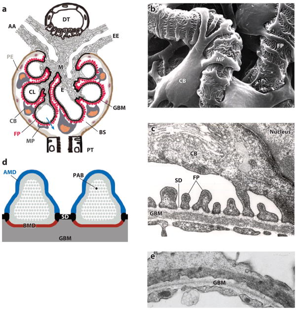
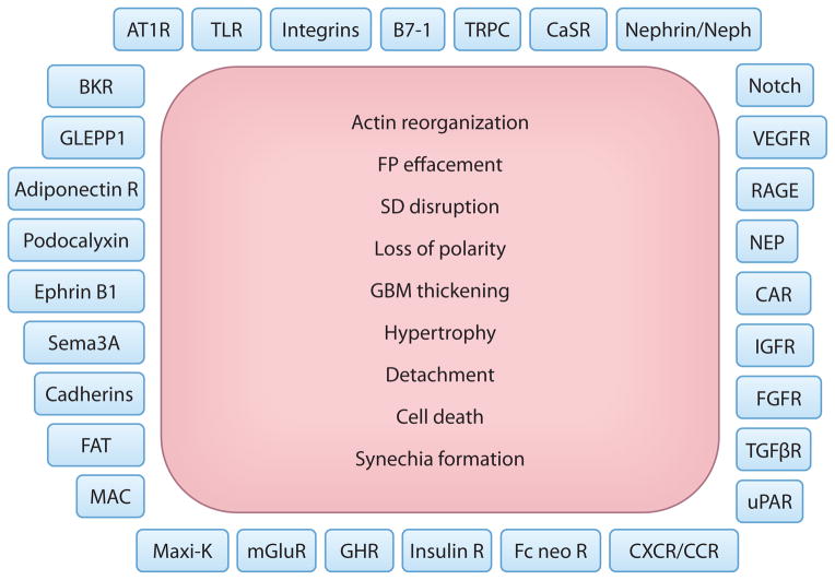
As an integral member of the filtration barrier in the kidney glomerulus, the podocyte is in a unique geographical position: It is exposed to chemical signals from the urinary space (Bowman's capsule), it receives and transmits chemical and mechanical signals to/from the glomerular basement membrane upon which it elaborates, and it receives chemical and mechanical signals from the vascular space with which it also communicates. As with every cell, the ability of the podocyte to receive signals from the surrounding environment and to translate them to the intracellular milieu is dependent largely on molecules residing on the cell membrane. These molecules are the first-line soldiers in the ongoing battle to sense the environment, to respond to friendly signals, and to defend against injurious foes. In this review, we take a membrane biologist's view of the podocyte, examining the many membrane receptors, channels, and other signaling molecules that have been implicated in podocyte biology. Although we attempt to be comprehensive, our goal is not to capture every membrane-mediated pathway but rather to emphasize that this approach may be fruitful in understanding the podocyte and its unique properties.


Figures

Figure 1
Figure 1
The function of podocytes is based on their intricate cell architecture. (a) A glomerulus contains a capillary tuft that receives primary structural support from the glomerular basement membrane (GBM). Glomerular endothelial cells (E) lining the capillary lumen (CL) and mesangial cells (M) are located on the blood side of the GBM, whereas podocyte foot processes (FP) cover the outer part of the GBM. Podocyte cell bodies (CB) and major processes (MP) float in Bowman’s space (BS) in primary urine. Along its route from the CL to BS (blue arrow), the plasma ultrafiltrate passes through the fenestrated endothelium, the GBM, and the filtration slits that are covered by the slit diaphragm (SD). AA, afferent arteriole; DT, distal tubule; EE, efferent arteriole; PE, parietal epithelium; PT, proximal tubule. (b) Scanning electron microscopy view from BS highlighting the intricate shape of podocytes. MP link the CB to FP, which interdigitate with FP of neighboring podocytes, thereby forming the filtration slits. (c) Transmission electron microscopy image of the filtration barrier consisting of fenestrated endothelium, GBM, and podocyte FP with the SD covering the filtration slits. (d) Podocyte FP are defined by three membrane domains: the apical membrane domain (AMD) (blue), the basal membrane domain (BMD) (red), and the SD (black). All three domains are connected to the underpinning actin cytoskeleton (gray) and with each other. PAB denotes a single parallel, contractile actin bundle containing α-actinin-4, myosin 9, and synaptopodin. (e) Under conditions of proteinuria, FP lose their normal interdigitating pattern and instead show effacement. A continuous sheet of cytoplasm filled with reorganized actin filaments is clearly visible.
Figure 2
Figure 2
Podocyte plasma membrane proteins and a canonical pattern of injury. Shown is an (incomplete) list of membrane proteins that have been implicated in the regulation of podocyte function in health and disease. Injured podocytes respond with a finite repertoire of changes, as depicted here. Our ability to repair the pathways initiated by the molecules on the podocyte plasma membrane to the precise cellular phenotypes listed here will provide not only novel insight but enormous opportunities for successful therapeutic interventions. Abbreviations: adiponectin R, adiponectin receptor; AT1R, angiotensin type 1 receptor; BKR, bradykinin receptor; CAR, Coxsackie and adenovirus receptor; CaSR, calcium-sensing receptor; CXCR/CCR, C-X-C/C-C chemokine receptor; FAT, protocadherin FAT1; Fc neo R, neonatal Fc receptor; FGFR, fibroblast growth factor receptor; FP, foot process; GBM, glomerular basement membrane; GHR, growth hormone receptor; GLEPP1, glomerular epithelial (podocyte) protein 1; IGFR, insulin growth factor receptor; insulin R, insulin receptor; MAC, membrane attack complex; Maxi-K, large-conductance calcium-activated potassium channel (also known as BK channel); mGluR, metabotropic glutamate receptor; NEP, neutral endopeptidase; RAGE, receptor for advanced glycation end products; SD, slit diaphragm; Sema3A, semaphorin-3A; TGF βR, transforming growth factor β receptor; TLR, Toll-like receptor; TRPC, transient receptor potential canonical; uPAR, urokinase receptor; VEGFR, vascular endothelial growth factor receptor.
Figure 3
Figure 3
Reversible and irreversible consequences of dysregulated podocyte signaling. (a) Dysregulated signaling at the plasma membrane may lead to reversible morphological changes. Therefore, proteinuria may arise, with or without foot process (FP) effacement (see text for details). However, if the upstream injurious signals are reversed, the cell morphology can revert back to physiological patterns. SD denotes slit diaphragm. (b) Persistence of podocyte injury is manifest in the activation of cellular processes that lead to irreversible changes such as loss of adhesion to the glomerular basement membrane (GBM), cell hypertrophy, transcriptional changes, disrupted metabolic pathways, autophagy, and cell cycle dysregulation. These irreversible changes can cause podocyte cell death or the detachment of podocytes from the GBM. Podocyte senescence may also be a manifestation of persistent injury, although less is known about this mechanism. The resulting loss of podocytes ultimately leads to irreversible glomerulosclerosis and kidney failure.
Figure 4
Figure 4
An example of pathways at the intersection of AT1R and TRPC signaling at the plasma membrane as they converge on synaptopodin and Rho GTPase signaling in the cytosol to effect critical cytoskeletal remodeling. Dashed arrows indicate indirect effects between molecules. Abbreviations: TLR, Toll-like receptor; AT1R, angiotensin type 1 receptor; TRPC6/5, transient receptor potential canonical channels 6 and 5; CathL, cathepsin L; PKA/CamKII, protein kinase A/calcium-calmodulin-dependent protein kinase II; CnA, calcineurin; PI3K, phosphoinositide 3 kinase; Synpo, synaptopodin; MR, mineralocorticoid receptor; FP, foot process.

References

    1. Mundel P, Kriz W. Structure and function of podocytes: an update. Anat Embryol. 1995;192:385–97. - PubMed
    1. Somlo S, Mundel P. Getting a foothold in nephrotic syndrome. Nat Genet. 2000;24:333–35. - PubMed
    1. Drenckhahn D, Franke RP. Ultrastructural organization of contractile and cytoskeletal proteins in glomerular podocytes of chicken, rat, and man. Lab Investig. 1988;59:673–82. - PubMed
    1. Kerjaschki D. Caught flat-footed: podocyte damage and the molecular bases of focal glomerulosclerosis. J Clin Investig. 2001;108:1583–87. - PMC - PubMed
    1. Takeda T, McQuistan T, Orlando RA, Farquhar MG. Loss of glomerular foot processes is associated with uncoupling of podocalyxin from the actin cytoskeleton. J Clin Investig. 2001;108:289–301. - PMC - PubMed

Publication types