Review
. 2011 Nov;51(5):313-4, 316, 318 passim.
doi: 10.2144/000113765.

Modern fluorescent proteins: from chromophore formation to novel intracellular applications

Affiliations
Review

Modern fluorescent proteins: from chromophore formation to novel intracellular applications

Olesya V Stepanenko et al. Biotechniques. 2011 Nov.

Abstract

The diverse biochemical and photophysical properties of fluorescent proteins (FPs) have enabled the generation of a growing palette of colors, providing unique opportunities for their use in a variety of modern biology applications. Modulation of these FP characteristics is achieved through diversity in both the structure of the chromophore as well as the contacts between the chromophore and the surrounding protein barrel. Here we review our current knowledge of blue, green, and red chromophore formation in permanently emitting FPs, photoactivatable FPs, and fluorescent timers. Progress in understanding the interplay between FP structure and function has allowed the engineering of FPs with many desirable features, and enabled recent advances in microscopy techniques such as super-resolution imaging of single molecules, imaging of protein dynamics, photochromic FRET, deep-tissue imaging, and multicolor two-photon microscopy in live animals.


Conflict of interest statement

Competing interests

The authors declare no competing interests.

Figures
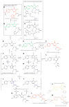
Figure 1
Figure 1. Chromophore formation pathways in the fluorescent proteins
A→B→C→D→E denotes the green chromophore formation pathway. A→B→H→J→K→L and A→B→C→G→I→K→L are two alternative pathways for red chromophore formation. L→N is the zFP538 chromophore formation pathway. L→M is the mOrange chromophore formation pathway. O→P is the light induced green-emitting chromophore formation pathway in the PA-GFP, PS-CFP and PS-CFP2 proteins. D→E→F is the oxidative redding pathway in the green FPs. Q→R is the light-induced chromophore transformation in the Kaede-like FPs.
Figure 2
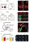
Figure 2. Advanced imaging techniques based on recently developed fluorescent proteins
(A) Schematic representation of STED microscopy (left panel) and the fine details of filopodia at the edge of the fixed HeLa cell revealed by STED microscopy (right panel, (68)). STED microscopy is based on the narrowing of an initial diffraction-limited excitation spot by applying a doughnut-shaped depletion laser pulse (STED pulse). The depletion results in an effective fluorescent spot of up to 20 nanometers in diameter. (B) Schematic representation of PALM microscopy (left panel) and application of two-color PALM microscopy for studies of colocalization of TfR tagged by PAmCherry1 and CLC tagged by PAGFP (right panel, (15)). PALM is based on a processing of the stack of images obtained by repeated activation of well-separated photoactivatable molecules, their localization using 2D Gaussian reconstruction, and subsequent bleaching. The resulting reconstituted image of the Gaussian centers provides a resolution of up to 20 nanometers. (C) Intravital imaging scheme relying on far-red/infrared FPs or far-red LSS-FPs and multiphoton excitation allowing subcellular resolution up to 150 μm in deep (left panel) and studies of tumor cell motility by 2P microscopy (right panel, (22)): the image of MTLn3 cells with stable coexpression of NLS-LSS-mKate1 (nucleus, red) and GalT-ECFP (Golgi, blue), and blood vessel labeled by FITC-dextran (green). (D) Photochromic FRET (pcFRET) scheme (left panel) and imaging of interaction between EGFR tagged with EYFP and Grb2 tagged with rsTagRFP in live HeLa cells using pcFRET (right panel, (18)) are shown. pcFRET is a technique for studying protein-protein interaction using the repeated switching on and off of the FRET process. The fluorescence emission of the FRET donor such as EYFP is modulated by the light-induced changes of absorbance spectrum of the rsTagRFP acceptor in its on and off states.

Similar articles

Cited by

References

    1. Wang Y, Shyy JY, Chien S. Fluorescence proteins, live-cell imaging, and mechanobiology: seeing is believing. Annu Rev Biomed Eng. 2008;10:1–38. - PubMed
    1. Pakhomov AA, Martynov VI. GFP family: structural insights into spectral tuning. Chem Biol. 2008;15:755–764. - PubMed
    1. Wu B, Piatkevich KD, Lionnet T, Singer RH, Verkhusha VV. Modern fluorescent proteins and imaging technologies to study gene expression, nuclear localization, and dynamics. Curr Opin Cell Biol 2011 - PMC - PubMed
    1. Frommer WB, Davidson MW, Campbell RE. Genetically encoded biosensors based on engineered fluorescent proteins. Chem Soc Rev. 2009;38:2833–2841. - PMC - PubMed
    1. Heim R, Prasher DC, Tsien RY. Wavelength mutations and posttranslational autoxidation of green fluorescent protein. Proc Natl Acad Sci U S A. 1994;91:12501–12504. - PMC - PubMed

Publication types

MeSH terms

Substances

LinkOut - more resources