. 2011 Nov 2;14(5):587-97.
doi: 10.1016/j.cmet.2011.09.012.

Dissociation of the glucose and lipid regulatory functions of FoxO1 by targeted knockin of acetylation-defective alleles in mice

Affiliations

Dissociation of the glucose and lipid regulatory functions of FoxO1 by targeted knockin of acetylation-defective alleles in mice

Alexander S Banks et al. Cell Metab. .

Abstract

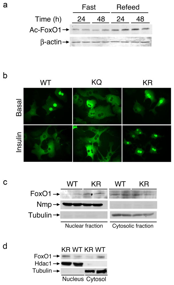
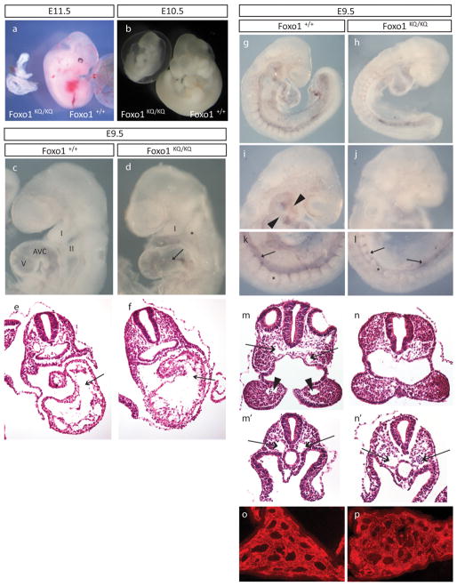
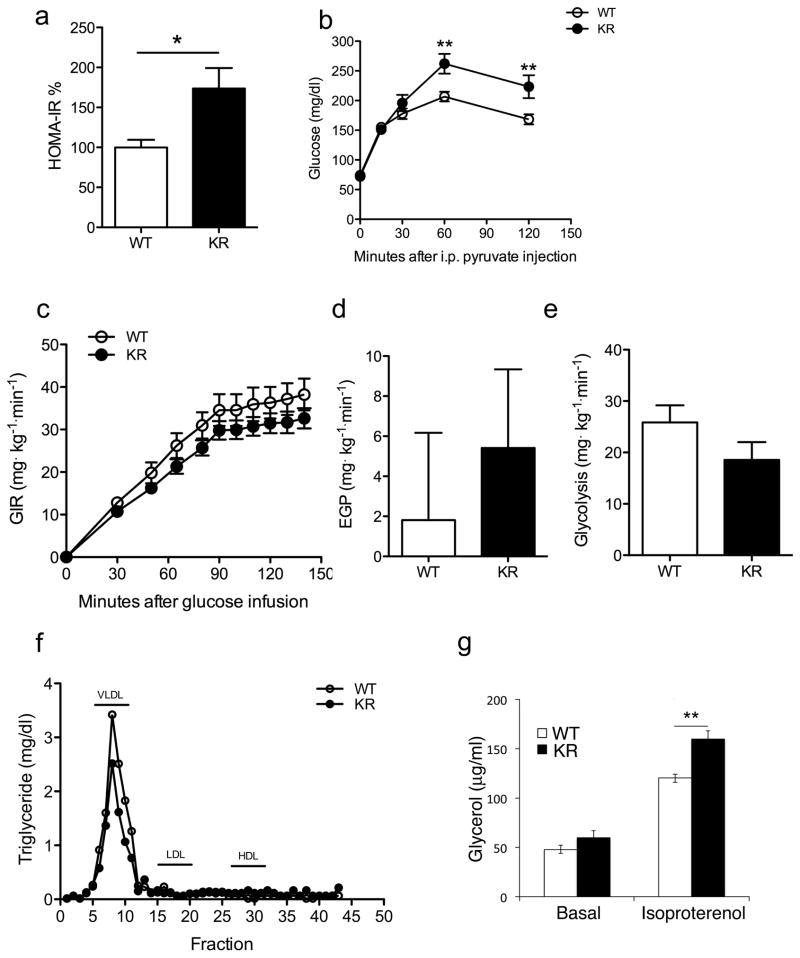
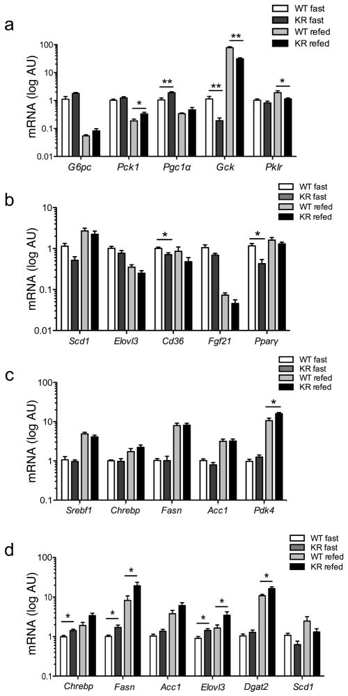
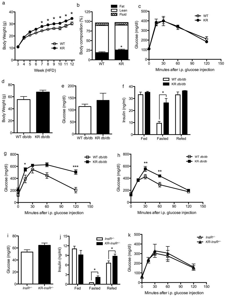
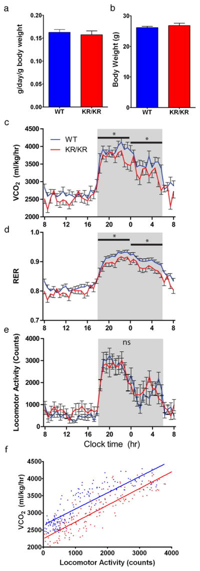
FoxO1 integrates multiple metabolic pathways. Nutrient levels modulate FoxO1 acetylation, but the functional consequences of this posttranslational modification are unclear. To answer this question, we generated mice bearing alleles that encode constitutively acetylated and acetylation-defective FoxO1 proteins. Homozygosity for an allele mimicking constitutive acetylation (Foxo1(KQ/KQ)) results in embryonic lethality due to cardiac and angiogenesis defects. In contrast, mice homozygous for a constitutively deacetylated Foxo1 allele (Foxo1(KR/KR)) display a unique metabolic phenotype of impaired insulin action on hepatic glucose metabolism but decreased plasma lipid levels and low respiratory quotient that are consistent with a state of preferential lipid usage. Moreover, Foxo1(KR/KR) mice show a dissociation between weight gain and insulin resistance in predisposing conditions (high fat diet, diabetes, and insulin receptor mutations), possibly due to decreased cytokine production in adipose tissue. Thus, acetylation inactivates FoxO1 during nutrient excess whereas deacetylation selectively potentiates FoxO1 activity, protecting against excessive catabolism during nutrient deprivation.


Figures

Figure 1
Figure 1. FoxO1 acetylation and localization
(A) Immunoblot analysis of FoxO1 acetyl-lysine content in epididymal adipose tissue extracts from mice subjected to fasting and re-refeeding. (B) Fluorescence microscopy of primary mouse hepatocytes transduced with FoxO1-GFP-WT, -KR or -KQ adenoviruses and maintained in serum-free medium (upper panel) or incubated with insulin (lower panel). We show representative images. (C–D) Immunoblotting analysis of FoxO1 content in nuclear and cytosolic fractions isolated from epididymal adipose tissue extracts of fasted (C) or refed (D) wild type (WT) or Foxo1KR/KR (KR) animals. We used nucleophosmin (NPM) or tubulin as markers of cellular fractions in (C) and Hdac1 or βactin in (D).
Figure 2
Figure 2. Embryonic lethality in mice homozygous for the Foxo1KQ/KQ mutation
(A–B) Microphotographs of Foxo1+/+ and Foxo1KQ/KQ embryos at E10.5 and E11.5. (C–D) Microphotographs of E9.5 embryos showing first (I) and second branchial arch (II), as well as atrioventricular canal (AVC) and ventricle (V). Arrow in D indicates the distended AVC in the mutant. (E–F) Histological sections of E9.5 embryos across the AVC (arrows) and ventricle. (G–L) Whole-mount PECAM-1 immunostaining with details of the aortic arch arteries in the head (arrowheads in I, missing in J) and the dorsal aorta (arrows) in the tail region (K–L) of a Foxo1+/+ (G, I, and K) and Foxo1KQ/KQ embryo (H, J, and L). Asterisks in k and l mark somites which are smaller in the mutant. (M–N) Histological sections through the first branchial arch showing dorsal aortae (arrows in M) and the first branchial arch artery (arrowheads in M). (M′-N′) Sections through more posterior regions showing paired dorsal aortae (arrows that are distended in the mutant (N′) compared with wild type (M′). (O–P) Vascular plexus in the yolk sac in Foxo1KQ/KQ and wild type embryos shown by immunostaining for PECAM.
Figure 3
Figure 3. Metabolic data in Foxo1KR/KR mice
(A) HOMA-IR values calculated from the data in Table S2. (B) Pyruvate tolerance tests in WT and Foxo1KR/KR mice. (C) Glucose infusion rates, (D) Endogenous glucose production under hyperinsulinemic conditions, and (E) Glycolysis during euglycemic hyperinsulinemic clamps. (F) Determination of TG content in lipoprotein fractions isolated by FPLC analysis. (G) Basal and isoproterenol-stimulated glycerol release from primary cultures of epididymal fat pads. n= ≥ 6 mice per group and each experiment. *= P<0.05; ** =P<0.01 by 2-way ANOVA. Data are presented as means ± SEM.
Figure 4
Figure 4. Gene expression analysis
(A)Hepatic expression of genes involved in glucose and (B, C) lipid metabolism in 2-month-old male mice. (D) Adipose expression of fatty acid and TG synthetic genes in the same cohort. n = ≥ 6 for each genotype. *=P<0.05, **=P<0.01 by Student’s t-test. Data are presented as means ± SEM.
Figure 5
Figure 5. Characterization of Foxo1KR/KR mice in dietary and genetic models of insulin resistance
(A) Body weight curves, (B) body composition, and (C) intraperitoneal glucose tolerance tests in WT and Foxo1KR/KR mice during high fat feeding. (D) Body weight, (E) 16-hr fasting glucose levels, (F) insulin levels in random fed, 16-hr fasted, and 4-hr re-fed, (G) glucose tolerance tests in male and (H) female db/db and Foxo1KR/KR;db/db mice. n=6–12 for each genotype and each experiment. *=P<0.05, **=P<0.01 by 2-way ANOVA. Data are presented as means ± SEM.
Figure 6
Figure 6. Energy homeostasis studies
(A) Food intake, (B) body weight, (C) energy expenditure as measured by CO2 produced (VCO2), (D) respiratory exchange ratio (CO2 produced/O2 consumed) (RER), (E) locomotor activity, and (F) metabolic efficiency plots in 2 month old WT and Foxo1KR/KR mice maintained on standard chow diet. n=8 for each genotype and each experiment. *=P<0.05 by 2-way ANOVA. Data are presented as means ± SEM.

References

    1. Accili D, Arden KC. FoxOs at the crossroads of cellular metabolism, differentiation, and transformation. Cell. 2004;117:421–426. - PubMed
    1. Accili D, Drago J, Lee EJ, Johnson MD, Cool MH, Salvatore P, Asico LD, Jose PA, Taylor SI, Westphal H. Early neonatal death in mice homozygous for a null allele of the insulin receptor gene. Nat Genet. 1996;12:106–109. - PubMed
    1. Allen-Jennings AE, Hartman MG, Kociba GJ, Hai T. The roles of atf3 in glucose homeostasis. a transgenic mouse model with liver dysfunction and defects in endocrine pancreas. J Biol Chem. 2001;276:29507–29514. - PubMed
    1. Altomonte J, Cong L, Harbaran S, Richter A, Xu J, Meseck M, Dong HH. Foxo1 mediates insulin action on apoC-III and triglyceride metabolism. J Clin Invest. 2004;114:1493–1503. - PMC - PubMed
    1. Banks AS, Kon N, Knight C, Matsumoto M, Gutierrez-Juarez R, Rossetti L, Gu W, Accili D. SirT1 gain of function increases energy efficiency and prevents diabetes in mice. Cell Metab. 2008;8:333–341. - PMC - PubMed

Publication types

MeSH terms