. 2011 Dec;121(12):4870-9.
doi: 10.1172/JCI40509. Epub 2011 Nov 7.

Connexins protect mouse pancreatic β cells against apoptosis

Affiliations

Connexins protect mouse pancreatic β cells against apoptosis

Philippe Klee et al. J Clin Invest. 2011 Dec.

Abstract

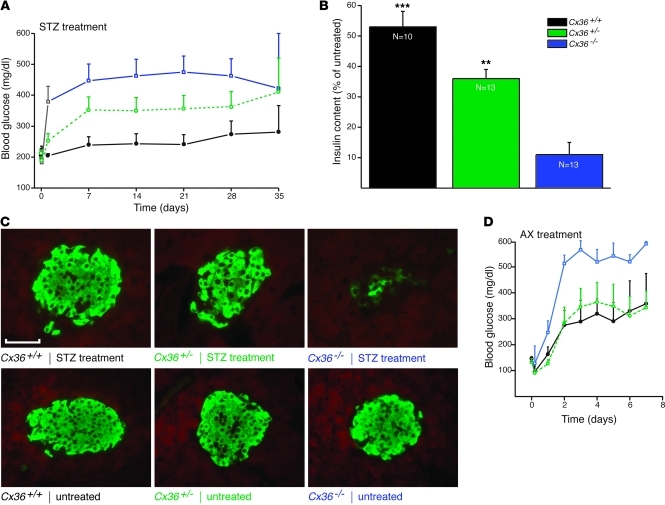
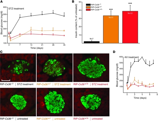
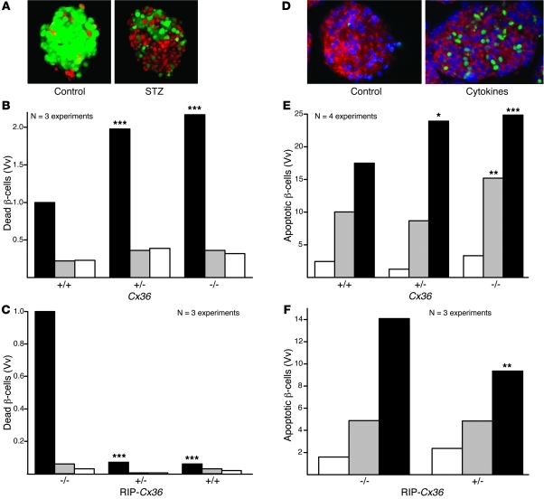
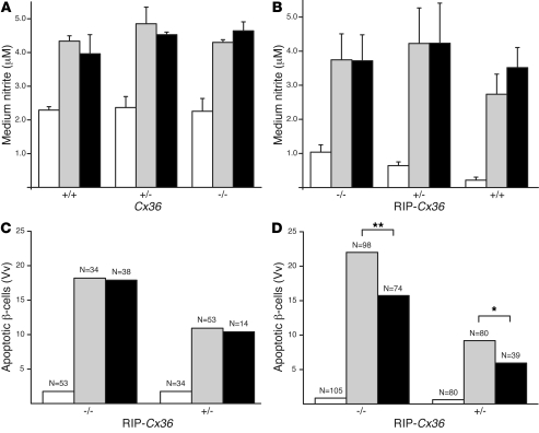
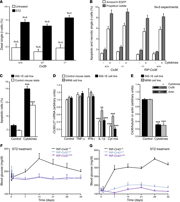
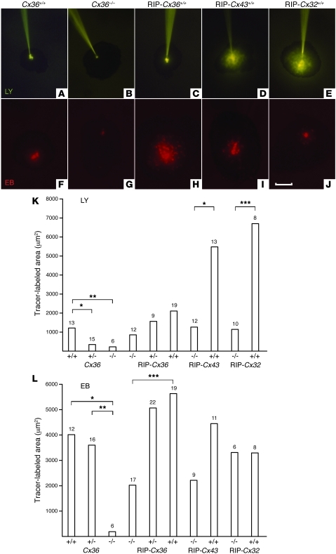
Type 1 diabetes develops when most insulin-producing β cells of the pancreas are killed by an autoimmune attack. The in vivo conditions modulating the sensitivity and resistance of β cells to this attack remain largely obscure. Here, we show that connexin 36 (Cx36), a trans-membrane protein that forms gap junctions between β cells in the pancreatic islets, protects mouse β cells against both cytotoxic drugs and cytokines that prevail in the islet environment at the onset of type 1 diabetes. We documented that this protection was at least partially dependent on intercellular communication, which Cx36 and other types of connexin channels establish within pancreatic islets. We further found that proinflammatory cytokines decreased expression of Cx36 and that experimental reduction or augmentation of Cx36 levels increased or decreased β cell apoptosis, respectively. Thus, we conclude that Cx36 is central to β cell protection from toxic insults.


Figures

Figure 1
Figure 1. Mice lacking Cx36 are sensitized to the toxic effects of STZ and AX.
(A) A single injection of 200 mg/kg STZ induced mild hyperglycemia in control Cx36+/+ mice (black; n = 9). This hyperglycemia was more pronounced in Cx36+/– (green; n = 14) and Cx36–/– (blue; n = 14) littermates, which expressed reduced and nil doses, respectively, of the Cx36 gene. (B) 5 weeks after STZ injection, pancreatic insulin content was highest in control Cx36+/+ mice, lowest in Cx36–/– mice, and intermediate in Cx36+/– mice. Values are expressed as percent of untreated; n as indicated. **P < 0.01, ***P < 0.001 versus Cx36–/–. (C) At the same time point, the proportion of surviving β cells, identified by insulin immunostaining, was highest in Cx36+/+, lowest in Cx36–/–, and intermediate in Cx36+/– mice. Compared with insulin staining of corresponding untreated mice, STZ-treated Cx36–/– mice revealed major loss of β cells. Scale bar: 120 μm. (D) A single injection of 60 mg/kg AX induced sustained hyperglycemia in Cx36+/+ (black; n = 4) and Cx36+/– (green; n = 4) mice. This hyperglycemia was significantly more pronounced in Cx36–/– littermates (blue; n = 5). All data are mean ± SEM.
Figure 2
Figure 2. Mice overexpressing islet Cx36 are protected against STZ and AX.
(A) STZ-induced hyperglycemia was observed in RIP-Cx36–/– mice (black; n = 15); in contrast, RIP-Cx36+/– (orange; n = 13) and RIP-Cx36+/+ (red; n = 11) mice did not become hyperglycemic for the duration of the experiment. (B) 5 weeks after STZ injection, residual insulin content was significantly higher in the pancreas of RIP-Cx36+/+ and RIP-Cx36+/– mice than in that of RIP-Cx36–/– littermates. Values are expressed as percent of the insulin content of untreated mice; n as indicated. ***P < 0.001 versus RIP-Cx36–/–. (C) The proportion of surviving insulin-containing β cells of RIP-Cx36+/+ and RIP-Cx36+/– mice was substantially higher than that of RIP-Cx36–/– littermates. As compared with insulin staining of the corresponding untreated islets, most β cells in RIP-Cx36+/– and RIP-Cx36+/+ islets were found to survive STZ treatment. Scale bar: 120 μm. (D) AX-induced hyperglycemia was also observed in RIP-Cx36–/– mice (black; n = 6), but not in RIP-Cx36+/– (orange; n = 4) and RIP-Cx36+/+ (red; n = 3) littermates. All data are mean ± SEM.
Figure 3
Figure 3. Cx36 protects β cells from Th1 cytokines.
(A) Islets isolated from C57BL/6 mice showed much more healthy cells (calcein stain; green) than dead cells (EB stain; red). The proportion of dead cells substantially increased after 24 hours exposure to STZ. (B) In islets of Cx36–/– mice, STZ (black) increased the volume density (Vv) of dead cells over that in untreated islets (white) and in islets exposed to citrate buffer (gray). The volume density of dead β cells was lower in the islets of STZ-treated Cx36+/+ mice than in those of Cx36+/– and Cx36–/– littermates. (C) Dead cell volume density was significantly higher in STZ-treated RIP-Cx36–/– islets than in islets of RIP-Cx36+/– and RIP-Cx36+/+ littermates. (D) TUNEL labeling showed that Cx36+/+ islets contained rare apoptotic cells (green). The number of apoptotic cells increased after 24 hours exposure to IL-1β, IFN-γ, and TNF-α. (E) Exposure of Cx36–/– islets to either IL-1β and IFN-γ (gray) or IL-1β, IFN-γ, and TNF-α (black) increased the volume density of apoptotic cells over that in untreated mice (white). Apoptosis was lower in the islets of cytokine-treated Cx36+/+ mice than in those of Cx36+/– and Cx36–/– littermates. (F) The volume density of apoptotic cells was higher in RIP-Cx36–/– islets than in islets of RIP-Cx36+/– littermates exposed to the 3 cytokines. Values are medians of the indicated number of experiments. *P < 0.05, **P < 0.01, ***P < 0.001 versus respective cognate control (Cx36+/+ or RIP-Cx36–/–).
Figure 4
Figure 4. Cytokines activate apoptotic pathways in β cells of mice from both Cx36 and RIP-Cx36 lines.
(A and B) A 24-hour exposure to either IL-1β plus IFN-γ (gray) or IL-1β, IFN-γ, and TNF-α (black) increased NO production to the same extent in islets isolated from mice of Cx36 (A) and RIP-Cx36 (B) lines. Values are mean ± SEM of the indicated number of independent experiments. (C) Exposure to 10 μM cyclosporin A (black) did not decrease the cytokine-induced (gray) apoptosis of RIP-Cx36 β cells. (D) β cell apoptosis increased over basal levels (white) in RIP-Cx36 islets isolated and exposed for 24 hours to a mix of IL-1β, IFN-γ, and TNF-α (gray). This increase was larger in islets isolated from RIP-Cx36–/– than in those of RIP-Cx36+/– littermates. In both groups, incubation in the presence of Z-VAD (black; added 1 hour prior to the cytokine and maintained throughout the incubation period) reduced the cytokine-induced apoptosis. Values are medians of the indicated number of islets. White bars throughout denote untreated (basal) levels. *P < 0.05, **P < 0.01.
Figure 5
Figure 5. β cell protection requires cell contact and Cx expression.
(A) The proportion of dead cells was similar in all control islet cell suspensions. STZ similarly increased this proportion in all groups. n as indicated. (B) Islet cells of control and homozygous mice of both Cx36 and RIP-Cx36 lines showed increased apoptosis (white) and necrosis (gray) after exposure to the cytokine mix of IL-1β, IFN-γ, and TNF-α. (C) The cytokine mix increased apoptosis of INS1E cells and control C57BL/6 islets. (D) The cytokine mix decreased Cx36 mRNA in mouse islets and in MIN6 and INS-1E cells. Values are expressed relative to L27 gene level. (E) The cytokine mix also decreased Cx36 in extracts of INS1E and MIN6 cells. Values are shown relative to the tubulin signal, normalized to control. Cx36 and actin Western blot immunolabeling, from which the quantitative data were generated, is also shown (inset). **P < 0.01, ***P < 0.001 versus corresponding control. (F) STZ injection induced hyperglycemia in RIP-Cx32–/– mice, but not in RIP-Cx32+/– and RIP-Cx32+/+ littermates. (G) RIP-Cx43–/– mice also became hyperglycemic after STZ injection, whereas RIP-Cx43+/– and RIP-Cx43+/+ littermates did not. Data are mean ± SEM of 4–6 experiments (A), of 3–5 experiments (BE), or of 3–12 mice (F and G).
Figure 6
Figure 6. Increased junctional coupling in islets resistant to β cell–toxic conditions.
(AE) Microinjection of islets with LY showed limited β cell coupling in islets of Cx36+/+ and RIP-Cx36+/+ mice (A and C), no coupling in islets of Cx36–/– mice (B), and greater coupling in islets of RIP-Cx43+/+ and RIP-Cx32+/+ mice (D and E). (FJ) Microinjection with EB showed sizable coupling in islets of Cx36+/+, RIP-Cx43+/+, and RIP-Cx32+/+ mice (F, I, and J), no coupling in islets of Cx36–/– mice (G), and greater coupling in islets of RIP-Cx36+/+ animals (H). Scale bar: 100 μm. (K and L) Coupling extent in islets from Cx36+/+, Cx36+/–, RIP-Cx43–/–, and RIP-Cx32–/– mice, as well as all mice from the RIP-Cx36 line, was larger after EB (L) than LY (K) injection. No coupling was observed with either tracer in Cx36–/– mice. Values show medians of the indicated number of islet microinjections. *P < 0.05, **P < 0.01, ***P < 0.001, median test.

References

    1. Bavamian S, et al. Islet-cell-to-cell communication as basis for normal insulin secretion. Diabetes Obes Metab. 2007;9 suppl 2:118–132. - PubMed
    1. Serre-Beinier V, et al. Cx36 preferentially connects beta-cells within pancreatic islets. Diabetes. 2000;49(5):727–734. doi: 10.2337/diabetes.49.5.727. - DOI - PubMed
    1. Serre-Beinier V, et al. Cx36 makes channels coupling human pancreatic beta-cells, and correlates with insulin expression. Hum Mol Genet. 2009;18(3):428–439. - PMC - PubMed
    1. Theis M, et al. Replacement by a lacZ reporter gene assigns mouse connexin36, 45 and 43 to distinct cell types in pancreatic islets. Exp Cell Res. 2004;294(1):18–29. - PubMed
    1. Harris AL, Locke D, eds.Connexin: A Guide . New York, New York, USA: Springer; 2009.

Publication types

MeSH terms