. 2012 Jan;177(1):32-9.
doi: 10.1016/j.jsb.2011.10.005. Epub 2011 Oct 25.

Formins filter modified actin subunits during processive elongation

Affiliations

Formins filter modified actin subunits during processive elongation

Qian Chen et al. J Struct Biol. 2012 Jan.

Abstract

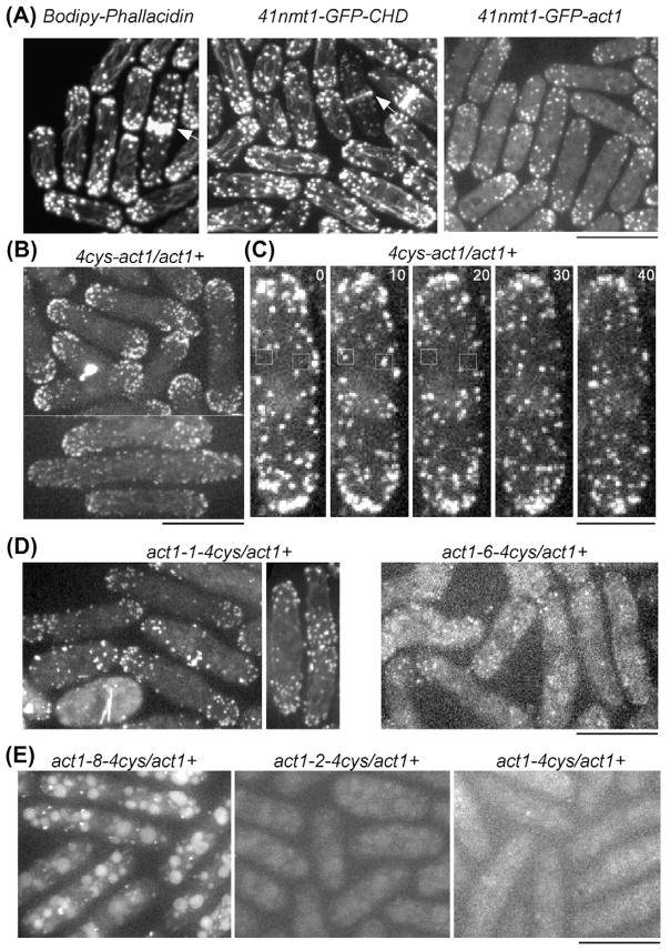
Fission yeast cells reject actin subunits tagged with a fluorescent protein from the cytokinetic contractile ring, so cytokinesis fails and the cells die when the native actin gene is replaced by GFP-actin. The lack of a fluorescent actin probe has prevented a detailed study of actin filament dynamics in contractile rings, and left open questions regarding the mechanism of cytokinesis. To incorporate fluorescent actin into the contractile ring to study its dynamics, we introduced the coding sequence for a tetracysteine motif (FLNCCPGCCMEP) at 10 locations in the fission yeast actin gene and expressed the mutant proteins from the native actin locus in diploid cells with wild-type actin on the other chromosome. We labeled these tagged actins inside live cells with the FlAsH reagent. Cells incorporated some of these labeled actins into actin patches at sites of endocytosis, where Arp2/3 complex nucleates all of the actin filaments. However, the cells did not incorporate any of the FlAsH-actins into the contractile ring. Therefore, formin Cdc12p rejects actin subunits with a tag of ~2 kDa, illustrating the stringent structural requirements for this formin to promote the elongation of actin filament barbed ends as it moves processively along the end of a growing filament.


Figures

Fig. 1
Fig. 1
Tetracysteine tag insertion sites on the actin monomer and filament. (A) Ribbon diagram of fission yeast actin (green ribbon) modeled (SWISS-MODEL (Arnold et al., 2006; Kiefer et al., 2009)) on the structure of budding yeast actin (PDB: 1YAG) with the tetracysteine tag insertion sites highlighted in yellow and denoted by black arrows. The three mutants, which bound FlAsH dye and localized to the actin patches in vivo, are labeled in blue. (B) Ribbon diagram of the actin filament model based on PDB: 2ZWH (Oda et al., 2009). The bound nucleotides are shown in red stick representation. The numbers indicate the subdomains of one actin subunit in the filament, which is oriented similar to the actin in (A). (C) Surface representation of the actin filament model in the various mutants, in the same orientation as (B) except where specifically denoted. The insertion sites are highlighted in yellow and the nucleotide is shown in red.
Fig. 2
Fig. 2
Comparison of methods to label actin filaments with fluorescent probes in fission yeast cells. Bars are 10 μM, except for that in (C) which is 5 μM. (A) Fluorescence micrographs of fission yeast cells with three different labeling strategies. Left, fixed cells stained with Bodipy-phallacidin. Middle, live cells expressing 41nmt1-GFP-CHD. Right, live cells expressing 41nmt1-GFP-act1 from the leu1 locus. Bodipy-phallacidin and GFP-CHD labeled all three cytoskeletal structures composed of actin filaments in the fission yeast cells, including actin patches, interphase actin cables and contractile rings (arrows). GFP-act1 localized only to the actin patches, not cables or contractile rings. (B–E) Fluorescence micrographs of live diploid cells expressing actin tagged with a tetracysteine peptide and labeled with FlAsH dye. (B and C) Cells expressing actin with an N-terminal tetracysteine tag and labeled with FlAsH. (B) FlAsH-4cys-act1 localized strongly to actin patches (upper) and very weakly to interphase actin cables (lower). (C) Time lapse fluorescence micrographs of cells showing the turnover of FlAsH-4cys-act1 in actin patches (squares). FlAsH-4cys-act1 appears and disappears in individual actin patches over about 10 s. Numbers are times in seconds. (D) Localization of two actin constructs with internal tetracysteine tags that localized to actin patches. Left, Act1-1-4cys labeled with FlAsH and localized to both actin patches and actin cables (right, weakly). Right, Act1-6-4cys labeled with FlAsH and localized to a small number of patches. (E) Actin constructs with tetracysteine tags that failed to localize to actin patches. Left, Act1-8-4cys, labeled with FlAsH but localized to aggregates and vacuoles. Middle, act1-2-4cys, labeled very weakly with FlAsH and concentrated in aggregates and vacuoles. Right, C-terminus tagged act1-4cys was distributed diffusely throughout the cells.
Fig. 3
Fig. 3
Models of tagged actins associated with an FH2 domain. (A) Space-filling model of the structure of one FH2 domain of budding yeast Bni1 cocrystallized with actin (PDB: 1Y64) (Otomo et al., 2005) showing the locations of sites tagged with GFP or tetracysteine. The surface rendering of the FH2 domain is blue and the ribbon diagram of actin is green. Red: locations of tetracysteine insertions. Blue: three subdomains of the FH2. A magenta arrow points to the insertion in act1-1 that could be labeled with FlAsH and incorporated into actin patches but not contractile rings. Black arrows point to insertions that could be labeled with FlAsH but were not incorporated into any actin cytoskeletal structures. Part of the DNase loop, N-terminus and C-terminus of the actin are missing in the structure. Therefore, act1-5, act1-6, 4cys-act1 and act1-4cys are not shown in this model. (B–D) show models of FH2 domains bound to actin filaments after adding a new subunit to the barbed end and before stepping onto this new subunit. (B) Cartoon of the barbed end of an actin filament (green), showing three subunits, associated with an FH2 dimer (one subunit in blue and the other in purple) as seen in (C) and (D). The actin subunit 1 (Actin 1) has the same orientation as actin in (A) with numbers (white) indicating its four subdomains. (C and D) Models of the FH2 domain bound to tetracysteine-tagged F-actin show how the tags may interfere with actin filament elongation. A single FH2 subunit (blue surface, 1Y64, (Otomo et al., 2005)) is bound to actin protomer 1 (as in B) of labeled filamentous actin (green ribbon, with insertions in yellow space filled representation PDB: 2ZWH (Oda et al., 2009)). (C) Act1-1-4cys tagged actin bound to FH2 (front view, center panel). The hairpin loop formed by the tetracysteine tag displays a clash with the knob region of FH2 (side view, left panel). Additionally, the insertion is also likely to impede the stepping of FH2 onto a subunit added to the barbed end (side view, right panel). (D) The Act1-6-4cys tagged actin bound to FH2 (front view, center panel). There are no obvious clashes between the tetracysteine insertion in the displayed conformation and bound FH2. However, the loop could prevent the movement of FH2 necessary for actin filament elongation (side view, left panel). The images were prepared with Pymol.

References

    1. Adams SR, Campbell RE, Gross LA, Martin BR, Walkup GK, et al. New biarsenical ligands and tetracysteine motifs for protein labeling in vitro and in vivo: synthesis and biological applications. J Am Chem Soc. 2002;124:6063–6076. - PubMed
    1. Aebi U, Smith PR, Isenberg G, Pollard TD. Structure of crystalline actin sheets. Nature. 1980;288:296–298. - PubMed
    1. Aizawa H, Sameshima M, Yahara I. A green fluorescent protein-actin fusion protein dominantly inhibits cytokinesis, cell spreading, and locomotion in Dictyostelium. Cell Struct Funct. 1997;22:335–345. - PubMed
    1. Arnold K, Bordoli L, Kopp J, Schwede T. The SWISS-MODEL workspace: a web-based environment for protein structure homology modelling. Bioinformatics. 2006;22:195–201. - PubMed
    1. Bahler J, Steever AB, Wheatley S, Wang Y, Pringle JR, et al. Role of polo kinase and Mid1p in determining the site of cell division in fission yeast. J Cell Biol. 1998;143:1603–1616. - PMC - PubMed

Publication types

MeSH terms

LinkOut - more resources