. 2011 Nov 1;25(21):2248-53.
doi: 10.1101/gad.173922.111.

Rejuvenating senescent and centenarian human cells by reprogramming through the pluripotent state

Affiliations

Rejuvenating senescent and centenarian human cells by reprogramming through the pluripotent state

Laure Lapasset et al. Genes Dev. .

Abstract

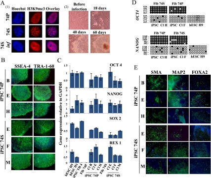
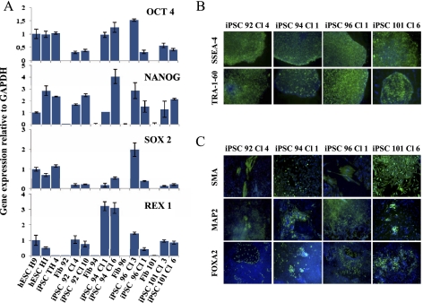
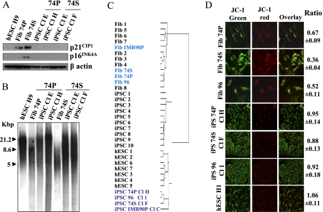
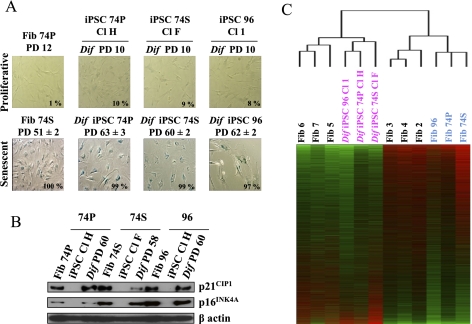
Direct reprogramming of somatic cells into induced pluripotent stem cells (iPSCs) provides a unique opportunity to derive patient-specific stem cells with potential applications in tissue replacement therapies and without the ethical concerns of human embryonic stem cells (hESCs). However, cellular senescence, which contributes to aging and restricted longevity, has been described as a barrier to the derivation of iPSCs. Here we demonstrate, using an optimized protocol, that cellular senescence is not a limit to reprogramming and that age-related cellular physiology is reversible. Thus, we show that our iPSCs generated from senescent and centenarian cells have reset telomere size, gene expression profiles, oxidative stress, and mitochondrial metabolism, and are indistinguishable from hESCs. Finally, we show that senescent and centenarian-derived pluripotent stem cells are able to redifferentiate into fully rejuvenated cells. These results provide new insights into iPSC technology and pave the way for regenerative medicine for aged patients.


Figures

Figure 1.
Figure 1.
Induction of pluripotency in proliferative and senescent 74-yr-old-derived cells. (A, panel 1) Detection of SAHF by indirect immunofluorescence of H3K9me3 (red) and Hoechst (blue) in proliferative (74P), senescent (74S), and transduced senescent 74-yr-old cells (74S inf) by the six factors. Seven days after transduction, no SAHF were detected. (Panel 2) Eighteen days after transduction, proliferation of infected 74S cells was observed (74S inf). Around day 40, distinct colonies were observed. Representative phase-contrast images are shown. (B) Immunodectection of surface markers TRA-1-60 and SSEA-4 on iPSCs colonies derived from 74S- and 74P-year-old cells (three independent clones). (C) Quantitative RT–PCR of expression levels for endogenous pluripotency factors in the iPSCs from 74P and 74S and their parental fibroblasts. H1 and H9 hESCs and iPSC TH 4 were used as controls. Transcript levels were normalized to GAPDH expression. Error bars indicate standard deviations from duplicate experiments. (D) Bisulfite sequencing analysis of OCT4 and NANOG promoter regions showing demethylation in iPSCs from 74P and 74S, as in H9 hESCs, compared with parental fibroblasts. Each column of circles for a given amplicon represents the methylation status of CpG dinucleotides in one clone for that region. Open circles are unmethylated CpGs and closed circles methylated ones. The left numbers of each column indicate CpG localization relative to the transcriptional start site. (E) In vitro differentiation experiments of iPSCs reveal their potential to generate cell derivatives of all three primary germ cell layers. Immunodetection of SMA, MAP2, and FOXA2 markers specific for endoderm, ectoderm, and mesoderm, respectively. Nuclei are stained with Hoechst (blue). Three independent clones are shown.
Figure 2.
Figure 2.
Induction of pluripotency in centenarian-derived cells. (A) Quantitative RT–PCR of expression levels of endogenous pluripotency factors in the indicated donor fibroblasts and iPSC lines. H1 and H9 hESCs as well as iPSC IMR90 TH Cl 4 cells were used as control. Transcript levels were normalized to GAPDH expression. Error bars indicate standard deviations from duplicate experiments. (B) Immunodetection of pluripotent cell surface markers TRA-1-60 and SSEA-4 on derived iPSCs colonies. (C) In vitro differentiation ability of iPSCs in the three primary germ cell layers revealed by immunodetection of SMA, MAP2, FOXA2 markers specific for endoderm, ectoderm, and mesoderm, respectively. Hoechst labeling was used for nuclear staining (blue).
Figure 3.
Figure 3.
Disappearance of senescence markers in senescent cell-derived iPSCs. (A) Decrease of p21CIP1 and p16INK4A protein level in iPSCs generated from 74P and 74S cells compared with parental fibroblasts and H9 hESCs, analyzed by Western blotting. β-Actin was used as a loading control. (B) TRF analysis of iPSC clones generated from 74P and 74S cells compared with their parental fibroblasts and H9 hESCs; TRF length is in kilobases (kb). (C) Nonsupervised hierarchical clustering of the global gene expression profiles in fibroblasts, iPSCs, hESCs, and 74- and 96-yr-old fibroblasts and their corresponding iPSCs. (D) Reprogramming of mitochondrial function in iPSCs derived from 74P and 74S and aged 96 cells compared with H1 hESCs, analyzed by JC-1 red/green fluorescence ratio measured by FACS. Red fluorescence indicates a normal membrane potential and green fluorescence indicates a membrane depolarization. A decreasing ratio measures the extent of mitochondrial dysfunction. Experiments were performed in triplicate (±SD for standard deviation).
Figure 4.
Figure 4.
Rejuvenated features of fibroblasts redifferentiated from iPSCs generated from senescent and aged fibroblasts with a six-factor-based reprogramming strategy. (A) SA-β-gal staining of proliferative (PD 10) and replicative senescence (PD 60–63) fibroblasts redifferentiated from iPSCs (PD 90). The percentage of positive cells is indicated in the inset. Experiments were performed in triplicate (±SD for standard deviation). (B) Restoration of p21CIP1 and p16INK4A protein expression in iPSC-derived fibroblasts is triggered during replicative senescence. Western blot analysis using β-actin as the loading control. (C) Nonsupervised hierarchical clustering with gene expression profiles of iPSCs redifferentiated into fibroblasts compared with their 74P-, 74S-, and 96-yr-old parental fibroblasts, postnatal fibroblasts, and embryonic fibroblasts differentiated from H1 hESCs.

References

    1. Alexander B, Coppola G, Perrault SD, Peura TT, Betts DH, King WA 2007. Telomere length status of somatic cell sheep clones and their offspring. Mol Reprod Dev 74: 1525–1537 - PubMed
    1. Armstrong L, Tilgner K, Saretzki G, Atkinson SP, Stojkovic M, Moreno R, Przyborski S, Lako M 2010. Human induced pluripotent stem cell lines show stress defense mechanisms and mitochondrial regulation similar to those of human embryonic stem cells. Stem Cells 28: 661–673 - PubMed
    1. Assou S, Le Carrour T, Tondeur S, Strom S, Gabelle A, Marty S, Nadal L, Pantesco V, Reme T, Hugnot JP, et al. 2007. A meta-analysis of human embryonic stem cells transcriptome integrated into a web-based expression atlas. Stem Cells 25: 961–973 - PMC - PubMed
    1. Banito A, Rashid ST, Acosta JC, Li S, Pereira CF, Geti I, Pinho S, Silva JC, Azuara V, Walsh M, et al. 2009. Senescence impairs successful reprogramming to pluripotent stem cells. Genes Dev 23: 2134–2139 - PMC - PubMed
    1. Campisi J 2011. Cellular senescence: putting the paradoxes in perspective. Curr Opin Genet Dev 21: 107–112 - PMC - PubMed

Publication types

Substances