. 2012 Jan 18;31(2):403-16.
doi: 10.1038/emboj.2011.385. Epub 2011 Nov 4.

Cdk1 promotes kinetochore bi-orientation and regulates Cdc20 expression during recovery from spindle checkpoint arrest

Affiliations

Cdk1 promotes kinetochore bi-orientation and regulates Cdc20 expression during recovery from spindle checkpoint arrest

Hongqing Liang et al. EMBO J. .

Abstract

The spindle assembly checkpoint (SAC), an evolutionarily conserved surveillance pathway, prevents chromosome segregation in response to conditions that disrupt the kinetochore-microtubule attachment. Removal of the checkpoint-activating stimulus initiates recovery during which spindle integrity is restored, kinetochores become bi-oriented, and cells initiate anaphase. Whether recovery ensues passively after the removal of checkpoint stimulus, or requires mediation by specific effectors remains uncertain. Here, we report two unrecognized functions of yeast Cdk1 required for efficient recovery from SAC-induced arrest. We show that Cdk1 promotes kinetochore bi-orientation during recovery by restraining premature spindle elongation thereby extinguishing SAC signalling. Moreover, Cdk1 is essential for sustaining the expression of Cdc20, an activator of the anaphase promoting complex/cyclosome (APC/C) required for anaphase progression. We suggest a model in which Cdk1 activity promotes recovery from SAC-induced mitotic arrest by regulating bi-orientation and APC/C activity. Our findings provide fresh insights into the regulation of mitosis and have implications for the therapeutic efficacy of anti-mitotic drugs.


Conflict of interest statement

The authors declare that they have no conflict of interest.

Figures

Figure 1
Figure 1
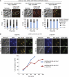
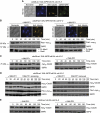
Recovery of cdc28-as1 cells from nocodazole-induced SAC arrest. (A) G1 synchronized cdc28-as1 slk19Δ cdc15-2 cells expressing Pds1–myc18 were released into nocodazole-containing medium at 30°C. Pds1–myc18, Mad1, G6PD levels and photomicrograph of cells arrested in nocodazole are shown. (B) Nocodazole-arrested cells as described in (A) were released in fresh medium with or without 1NM-PP1. Pds1–myc18, Mad1, Clb2–HA3, G6PD levels, DNA content and spindles were monitored. Bar scale: 5 μm. (C) Graph showing proportion of cells that recovered from nocodazole arrest as determined by their entry into anaphase. (D) cdc28-as1 slk19Δ cdc15-2 cells arrested with nocodazole treatment were released into medium with or without 1NM-PP1. Photomicrograph shows Spc42–RFP (marking the SPBs) and CENV–GFP staining at 120 min. The graph shows the bi-orientation kinetics. In all, 100–200 cells were analysed for each sample. Each experiment was performed at least twice and one representative experiment is shown here. Bar scale: 5 μm. Figure source data can be found in Supplementary data.
Figure 2
Figure 2
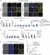
Cdk1 inhibition does not affect kinetochore capturing. (A) cdc28-as1 GAL-CENIII–GFP SPC42–RFP MET-CDC20 cells were incubated in nocodazole-containing YEP+Raff+Gal+Met medium and then released into three different media conditions: (1) YEP+Gal+Met+1NM-PP1 medium, (2) YEP+Glu+Met+1NM-PP1 medium and (3) YEP+Glu+Met−1NM-PP1 medium. Photomicrographs depict the positions of CENIII–GFP and Spc42–RFP 30 min after release. Bar scale: 5 μm. Bottom panels: bar charts showing proportion of cells with distances between CENIII–GFP and Spc42–RFP after release from nocodazole arrest. Approximately 100 cells were analysed for each sample. (B) Bi-orientation in cdc28-as1 slk19Δ cdc15-2 cells after release from HU arrest in absence or presence of 1NM-PP1. Photomicrographs show the state of nuclear division, spindle staining and CENV–GFP signal 60 and 120 min after release from HU arrest. Bar scale: 5 μm. Graph depicts bi-orientation kinetics after the release. Experiments were performed twice and >200 cells were analysed for each sample.
Figure 3
Figure 3
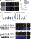
Spindle length and bi-orientation in cdc28-as1 cells after release from nocodazole. (A) cdc28-as1 slk19Δ cdc15-2 cells were arrested in nocodazole and subsequently released into 1NM-PP1 containing medium for 120 min. 1NM-PP1 was then removed to allow recovery. Photomicrographs show the state of nuclear division, spindle staining and CENV–GFP in 1NM-PP1 medium (left panel) and after removal of 1NM-PP1 (right panel). Bar scale: 5 μm. (B) Nocodazole-arrested cdc28-as1 slk19Δ cdc15-2 cells were released in the absence or presence of 1NM-PP1. One half of 1NM-PP1-treated culture was further allowed to recover in 1NM-PP1-free medium to restore Cdk1 activity. The bar charts represent percentage of cells with different spindle lengths. (CE) cdc28-as1 slk19Δ cdc15-2 and cdc28-as1 cin8Δ slk19Δ cdc15-2 cells were released from nocodazole arrest into medium containing 1NM-PP1. (C) Graphs shows the proportion of cells with different spindle lengths. (D) Spindle staining and CENV–GFP bi-orientation. Bar scale: 5 μm. (E) Kinetics of CENV–GFP bi-orientation. Approximately 100 cells were analysed for every sample in each experiment.
Figure 4
Figure 4
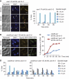
Ase1 deficiency prevents premature spindle extension in cdc28-as1 cells after nocodazole release. (AC) cdc28-as1 slk19Δ cdc15-2 and cdc28-as1 ase1Δ slk19Δ cdc15-2 cells were released from nocodazole into medium containing 1NM-PP1. (A) Spindle staining and CENV–GFP 20 and 80 min after the release from nocodazole. Bar scale: 5 μm. (B) Bi-orientation kinetics. (C) Graphs showing percentage of cells with different spindle lengths. Approximately 100 cells were analysed for every sample in the experiment. (D) Ase1 phosphorylation status correlated with spindle length dynamics in cdc28-as1 cells. cdc28-as1 slk19Δ cdc15-2 expressing Ase1–HA cells were released from nocodazole in the absence or presence of 1NM-PP1. Western blot shows Ase1–HA immunoprecipitated from whole-cell lysate, Ase1–HA immunoprecipitates treated with CIP (+CIP) and G6PD from whole-cell lysate. The state of spindles is shown in the right panel. Bar scale: 5 μm. Figure source data can be found in Supplementary data.
Figure 5
Figure 5
Effect of phosphomimetic mutants of Ase1 on spindle elongation and bi-orientation. (A) Spindle staining in cdc28-as1 slk19Δ cdc15-2 ase1-7A cells after release from nocodazole arrest without Cdk1 inhibition. White arrows indicate cells with unequal nuclear division. Bar chart depicts percentage of cells with different spindle lengths. (B) Spc42–RFP and CENV–GFP signals in cdc28-as1 slk19Δ cdc15-2 and cdc28-as1 ase1-7D slk19Δ cdc15-2 cells after release from nocodazole arrest in the presence of 1NM-PP1. Bar scale: 5 μm. (C) Bi-orientation kinetics in cells described in (B). (D) The proportion of cells with various spindle lengths in the experiment described in (B). Approximately 100 cells were analysed for every sample in each experiment.
Figure 6
Figure 6
Recovery of cdc28-as1 cells from GAL-MPS1-induced SAC arrest. (A) cdc28-as1 slk19Δ cdc15-2 GAL-MPS1 cells were arrested in metaphase by growing in YEP+Raff+Gal medium. Cells show short spindles and bi-orientated CENV–GFP. Bar scale: 5 μm. (B) Cells arrested in (A) were released into glucose medium with or without 1NM-PP1. Spindles and Pds1–myc18, Mad1, G6PD levels were monitored. (C) Levels of Cdc20 protein in cells described in (B). (D) cdc28-as1 slk19Δ cdc15-2 GAL-MPS1 cells arrested in galactose medium were released into glucose medium containing 1NM-PP1. The culture was divided into two portions: while one half remained in 1NM-PP1 medium; the cells from the other half were released into 1NM-PP1-free medium. Myc8–Cdc20, Pds1–myc18 and G6PD levels were monitored. (E) cdc28-as1 slk19Δ cdc15-2 cells were arrested in nocodazole and then released into the medium with or without 1NM-PP1. Cdc20 levels were determined by western blotting. Figure source data can be found in Supplementary data.
Figure 7
Figure 7
Cdk1 kinase activity regulates CDC20 transcription in mitosis. (A) cdc28-as1 cells with GAL-myc4–CDC20 were arrested in nocodazole-containing raffinose medium. Cells were induced to express Cdc20 for 60 min in YEP+Raff+Gal medium and then shifted to YEP+Glu+cycloheximide medium in absence or presence of 1NM-PP1. The status of myc-tagged Cdc20 protein was monitored by western blotting. (B) cdc28-as1 slk19Δ cdc15-2 cells were arrested in nocodazole. The culture was divided into two halves and cells were re-suspended in medium with or without 1NM-PP1. For cells with 1NM-PP1, 1NM-PP1 was removed from one half of the culture after 60 min to allow restoration of Cdk1 kinase activity. (C) Endogenous CDC20 in cdc28-as1 slk19Δ cdc15-2 cells was replaced by GAL-CDC20. Cells were arrested in nocodazole+Gal medium and then re-suspended in nocodazole-containing +Gal medium without or with 1NM-PP1. (D) CDC20 mRNA levels in cdc28-as1 slk19Δ cdc15-2 and cdc28-as1 yox1Δ slk19Δ cdc15-2 cells arrested in nocodazole and subsequently treated with 1NM-PP1. (E) Cdc20 protein levels in cdc28-as1 slk19Δ cdc15-2 and cdc28-as1 yox1Δ slk19Δ cdc15-2 cells arrested in nocodazole and then treated with 1NM-PP1. (F) Nocodazole-arrested cdc28-as1 slk19Δ cdc15-2 cells were divided into two portions. Cells from one half of the culture were re-suspended in nocodazole-containing medium without 1NM-PP1 and from the other half with 1NM-PP1. Yox1–HA6 protein level (left panel) and YOX1 mRNA level (right panel) are shown. Figure source data can be found in Supplementary data.

References

    1. Amon A (1999) The spindle checkpoint. Curr Opin Genet Dev 9: 69–75 - PubMed
    1. Bachant J, Jessen SR, Kavanaugh SE, Fielding CS (2005) The yeast S phase checkpoint enables replicating chromosomes to bi-orient and restrain spindle extension during S phase distress. J Cell Biol 168: 999–1012 - PMC - PubMed
    1. Biggins RH, Murray AW (2001) The budding yeast protein kinase Ipl1/Aurora allows the absence of tension to activate spindle checkpoint. Genes Dev 15: 3118–3129 - PMC - PubMed
    1. Bishop AC, Ubersax JA, Petsch DT, Matheos DP, Gray NS, Blethrow J, Shimizu E, Tsien JZ, Schultz PG, Rose MD, Wood JL, Morgan DO, Shokat KM (2000) A chemical switch for inhibitor-sensitive alleles of any protein kinase. Nature 407: 395–401 - PubMed
    1. Cross F, Tinkelenberg AH (1991) A positive feedback loop controlling CLN1 and CLN2 gene expression at the START of yeast cell cycle. Cell 65: 875–883 - PubMed

Publication types

MeSH terms

Substances