. 2012 Jan 15;361(2):301-12.
doi: 10.1016/j.ydbio.2011.10.027. Epub 2011 Oct 26.

SOHLH1 and SOHLH2 coordinate spermatogonial differentiation

Affiliations

SOHLH1 and SOHLH2 coordinate spermatogonial differentiation

Hitomi Suzuki et al. Dev Biol. .

Abstract

Spermatogonial self-renewal and differentiation are essential for male fertility and reproduction. We discovered that germ cell specific genes Sohlh1 and Sohlh2, encode basic helix-loop-helix (bHLH) transcriptional regulators that are essential in spermatogonial differentiation. Sohlh1 and Sohlh2 individual mouse knockouts show remarkably similar phenotypes. Here we show that SOHLH1 and SOHLH2 proteins are co-expressed in the entire spermatogonial population except in the GFRA1(+) spermatogonia, which includes spermatogonial stem cells (SSCs). SOHLH1 and SOHLH2 are expressed in both KIT negative and KIT positive spermatogonia, and overlap Ngn3/EGFP and SOX3 expression. SOHLH1 and SOHLH2 heterodimerize with each other in vivo, as well as homodimerize. The Sohlh1/Sohlh2 double mutant phenocopies single mutants, i.e., spermatogonia continue to proliferate but do not differentiate properly. Further analysis revealed that GFRA1(+) population was increased, while meiosis commenced prematurely in both single and double knockouts. Sohlh1 and Sohlh2 double deficiency has a synergistic effect on gene expression patterns as compared to the single knockouts. SOHLH proteins affect spermatogonial development by directly regulating Gfra1, Sox3 and Kit gene expression. SOHLH1 and SOHLH2 suppress genes involved in SSC maintenance, and induce genes important for spermatogonial differentiation.


Figures

Figure 1
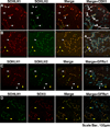
Figure 1. SOHLH1 and SOHLH2 are co-expressed in GFRA1 undifferentiated spermatogonia
Whole mount immunostaining of seminiferous tubule of adult testes (7-week-old) during stages V–VIII. (A–C) Tubules were immunostained with anti-SOHLH1 (red), anti-SOHLH2 (green) and anti-CDH1(cyan) or anti-GFRA1(cyan). White arrowheads indicate spermatogonia in which SOHLH1 and SOHLH2 were co-expressed with CDH1 or GFRA1. Yellow arrowheads indicate spermatogonia devoid of SOHLH1 and SOHLH2, but positive for CDH1 or GFRA1. (D) Tubules were immunostained with anti-SOHLH1 (green), anti-SOX3 (red) and anti-GFRA1 (cyan). Yellow arrowheads indicate SOHLH1 and SOX3 negative, GFRA1 positive spermatogonia. SOX3 was co-expressed with SOHLH1 in majority of undifferentiated spermatogonia. Scale bar, 100µm.
Figure 2
Figure 2. SOHLH1 and SOHLH2 are co-expressed in KIT-positive spermatogonia
Tubules were immunostained with anti-SOHLH1 (green), anti-KIT (cyan) and anti-ZBTB16 (red). Representative examples of the staining patterns that were observed for each group of epithelial stages are shown. (A–C) stages I–VI; (C and D) stages V–VIII; (E and F) stages IX–XI; (F, G and A) stages X–III. There is overlap of some stages among groups. The Roman numerals on the right side of the panels indicate the estimated stages of epithelial cycle from these observations (see supplementary Materials and Methods). White arrowheads indicate the SOHLH1+/ZBTB16+/KIT undifferentiated spermatogonia. Yellow arrowheads indicate the SOHLH1/ZBTB16+/KIT undifferentiated spermatogonia. Magenta arrowheads indicate the SOHLH1+/ZBTB16+/KIT+ differentiating spermatogonia. SOHLH1 is expressed in the KIT+ differentiating spermatogonia but not in the spermatocytes. The same pattern of expression was observed for SOHLH2 (data not shown).
Figure 3
Figure 3. SOHLH1 and SOHLH2 can form homodimers and heterodimers
(A) Co-immunoprecipitation analyses with guinea pig anti-SOHLH1 (α-S1) and guinea pig anti-SOHLH2 (α-S2) antibodies using 1-week-old testes extracts from wild type, Sohlh1−/− and Sohlh2−/− mice. Each antibody used for western blot analysis (W.B.) is indicated. SOHLH2 was co-immunoprecipitated with SOHLH1 and vice versa (I.P.), indicating that SOHLH1 and SOHLH2 heterodimerize. Input: pre-immunoprecipitation testes lysate (1%), IgG: normal rabbit IgG or normal guinea pig IgG. (B) Co-immunoprecipitation analyses with anti-FLAG antibody using 293T cells over-expressing MYC- or FLAG-tagged SOHLH1 or SOHLH2 proteins. Each combination of transfected vectors and each antibody used for western blot analysis are indicated. The lanes highlighted by yellow indicate that SOHLH1 and SOHLH2 can form homodimers. 3xMYC-SOHLH1 co-immunoprecipitated with 3xFLAG-SOHLH1, and 3xMYC-SOHLH2 co-immunoprecipitated with 3xFLAG-SOHLH2, indicating that both SOHLH1 and SOHLH2 are capable of forming homodimers. Input: pre-immunoprecipitation cell lysate (1%), Mock: pcDNA-3.1(+)-3FLAG.
Figure 4
Figure 4. SOHLH1 and SOHLH2 double knockouts arrest spermatogonial differentiation and induce precocious meiosis
(A–L) Histological analyses of wild type (A, E and I), Sohlh1−/− (B, F and J), Sohlh2−/− (C, G and K) and Sohlh1−/−/Sohlh2−/− (D, H and L) testes. Samples were prepared at one week (A–D), 3 weeks (E–H) and 5 weeks (I–L) after birth. (A–D) Each section was stained with Periodic Acid Schiff-Hematoxylin (PAS-H). (E–H) Each section was immunostained with anti-ZBTB16/PLZF antibody and counterstained with hematoxylin. (I–L) Samples were prepared 3 hours after BrdU injection to mice. Each section was immunostained with anti-BrdU antibody and counterstained with hematoxylin. (M–P) Immunofluorescence of wild type (M), Sohlh1−/− (N), Sohlh2−/− (O) and Sohlh−/−/Sohlh2−/− (P) testes at 1 week. Each section was immunostained with anti-HORMAD1 (green) and anti-CDH1 (red), and counterstained with DAPI (blue). White arrowheads indicate meiotic cells. Inset in (O) panel shows higher magnification of cells showed thread-like chromosome condensations. Single and double knockout testes germ cells entered meiosis precociously. Scale bars, 100µm.
Figure 5
Figure 5. SOHLH1 and SOHLH2 double deficiency has a synergistic effect on gene expression
(A and B) Gene expression array data comparing single and double Sohlh1/Sohlh2 mutant mice. (A) Hierarchical clustering analysis. Each row corresponds to a specific genotype shown at the bottom of the column, and each column corresponds to the relative expression profile of each genes. The color scheme reflects the expression level against the median of 4 genotypes, and the range bar is shown (0 is median). Sohlh1−/− (S1−/−) and Sohlh2−/− (S2−/−) single mutants showed overall a similar pattern of gene expression overall, while the Sohlh1−/−/Sohlh2−/− double mutant (dKO) showed a remarkably different pattern from either the wild type (WT) or single knockouts. (B) Venn diagram of gene expression changes in Sohlh1−/− (green), Sohlh2−/− (blue) and Sohlh1−/−/Sohlh2−/− (dKO, red). The intersection of genotypes indicates the number of genes where expression changed in two or all Sohlh mutants. Upward arrows indicate numbers of genes with enhanced expression, and downward arrows indicate reduced expression. Nearly 4 times as many genes were affected in the double mutant as compared to the single mutants. (C–F) Quantitative real-time PCR analysis on 1-week-old testis for genes differentially expressed between wild type (white), Sohlh1−/− (light gray), Sohlh2−/− (dark gray) and Sohlh1−/−/Sohlh 2−/− (dKO, black). Each gene is indicated at the bottom of the respective column. Genes involved in: (C) spermatogonial differentiation, (D) spermatogonial stem cell maintenance, (E) mRNA translational regulation, and (F) meiosis and epigenetics were evaluated. Data were normalized against Gapdh expression and are given as the mean relative quantity, with error bars representing the SEM. Student t-test was used to calculate P values. Significance was shown as a single asterisk when P < 0.05 against wild type. A double asterisk was used to show when double knockout expression significantly differed (P<0.05) not only against the wild type but also against Sohlh1 and Sohlh2 single knockouts (Spo11, Rec8 and Tia1).
Figure 6
Figure 6. SOHLH1 and SOHLH2 bind chromatin upstream of genes essential for SSC maintenance and spermatogonial differentiation
Chromatin immunoprecipitation (ChIP) assay. Anti-SOHLH1(αS1) and anti-SOHLH2 (αS2) antibodies precipitate genomic DNA containing conserved E-boxes surrounding Gfra1 (A), Sohlh1 (B), Sohlh2 (C) Sox3 (D) and Kit (E) genes. Kit position −397/388 relative to the transcriptional initiation site is an E-box that was not immunoprecipitated significantly with anti-SOHLH1 antibodies (E), and Kit position −998 is an E-box that was not immmunopreicipitated with both anti-SOHLH1 and anti-SOHLH2 antibodies. One-week-old testes from the wild type (WT), Sohlh1−/− (S1−/−) and Sohlh2−/− (S2−/−) mice were used for ChIP. “Input” is the PCR product from chromatin pellets before immunoprecipitation (1% of the total volume used for IP). Numbers at the left of each figure indicate the E-box location from the transcription start site.
Figure 7
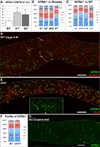
Figure 7. SOHLH1 and SOHLH2 are involved in differentiation from the most primitive set of undifferentiating spermatogonia
(A) The graph shows the percentage of GFRA1+ spermatogonia in the ZBTB16+ population in one-week-old testes of wild type (WT), Sohlh1−/− (S1−/−) and Sohlh2−/− (S2−/−) mice. Error bars represent the SEM. Student t-test was used to calculate P values. A single asterisk signifies a statistically significant (P < 0.05) increase in the number of GFRA1+/ ZBTB16+ spermatogonia as compared to the wild type. (B and C) Morphological profile of GFRA1+ spermatogonia in wild type and mutant testes. As −1 (blue), Apr −2 (red), chain of 3 cells −3 (green), Aal-4 −4 (purple) and chain of more than 4 cells -M (turquoise). Numbers displayed within each colored region indicate the percentage of the GFRA1+ population in tubules. The percentages for the green and turquoise regions in C are not shown. (B) GFRA1+ profile by spermatogonial morphology by genotype: Sohlh1−/− (S1−/−), Sohlh2−/− (S2−/−), Sohlh1−/−/Sohlh2−/− (dKO) and wild type (WT). (C) GFRA1+ profile by spermatogonial morphology at different epithelial stages in the wild type testes. The stages are indicated at the bottom of its respective bar. (D and E) Whole mount immunostaining of testes seminiferous tubules. Dotted lines indicate the outline of the tubule. (D) Wild type tubules during stages X-III were immunostained with anti-GFRA1 (green), and anti-SOHLH1 (red). (E) Sohlh1−/−/Sohlh2−/− (dKO) tubule was immunostained with anti-GFRA1 (green), and anti-DDX4/MVH (red). The yellow arrowhead indicates an unusually large cluster of GFRA1 spermatogonia. White dots and arrowheads in the inset show a long GFRA1+ chain of more than 8 spermatogonia. Asterisks in D and E indicate non-specific staining of interstitial cells. (F) Graphic representation of the distribution of morphological profiles for GFRA1+ spermatogonia in the wild type (WT) and experimentally induced cryptorchid testes (CRYP) in wild type mice. As −1 (blue), Apr −2 (red), 3 cell chains −3 (green), Aal-4 −4 (purple) and chains with more than 4 cells -M (turquoise). Numbers displayed within the blue, red and purple regions indicate the percentages of each type of cell in the total GFRA1+ population. There was not a significant difference between wild type (WT) and cryptorchid testis (CRYP). (G) Whole mount immunostaining of seminiferous tubules from experimentally induced cryptorchid wild type testes. Tubules were immunostained with anti-GFRA1 antibody (green). Notice the lack of long GFRA1+ chains, present in Sohlh mutants. Dotted lines indicate outline of the tubule. Scale bar, 100µm.
Figure 8
Figure 8. SOHLH1 and SOHLH2 regulation of spermatogonial development
In this figure, DMRT1 is shown as a direct regulator of SOHLH1. Current data do not support direct regulation of SOHLH2 by DMRT1 (Matson et al., 2010). DMRT1 is unlikely to be the sole regulator of SOHLH1 as spermatogonial differentiation proceeds in the DMRT1 conditional mutants (Matson et al., 2010). SOHLH1 and SOHLH2 bind the promoters of key spermatogonial molecules Gfra1, Sox3, Sohlh1, Sohlh2 and Kit. Solid lines indicate presumed direct transcriptional control, and dashed lines with question marks indicate undefined regulatory interactions.

References

    1. Anderson EL, Baltus AE, Roepers-Gajadien HL, Hassold TJ, de Rooij DG, van Pelt AM, Page DC. Stra8 and its inducer, retinoic acid, regulate meiotic initiation in both spermatogenesis and oogenesis in mice. Proc Natl Acad Sci U S A. 2008;105:14976–14980. - PMC - PubMed
    1. Baleato RM, Aitken RJ, Roman SD. Vitamin A regulation of BMP4 expression in the male germ line. Dev Biol. 2005;286:78–90. - PubMed
    1. Ballow D, Meistrich ML, Matzuk M, Rajkovic A. Sohlh1 is essential for spermatogonial differentiation. Dev Biol. 2006a;294:161–167. - PubMed
    1. Ballow DJ, Xin Y, Choi Y, Pangas SA, Rajkovic A. Sohlh2 is a germ cell-specific bHLH transcription factor. Gene Expr Patterns. 2006b;6:1014–1018. - PubMed
    1. Baltus AE, Menke DB, Hu YC, Goodheart ML, Carpenter AE, de Rooij DG, Page DC. In germ cells of mouse embryonic ovaries, the decision to enter meiosis precedes premeiotic DNA replication. Nat Genet. 2006;38:1430–1434. - PubMed

Publication types

MeSH terms

Substances