. 2011 Dec;93(6):937-46.
doi: 10.1016/j.exer.2011.10.011. Epub 2011 Nov 2.

A novel melano-lysosome in the retinal epithelium of rhesus monkeys

Affiliations

A novel melano-lysosome in the retinal epithelium of rhesus monkeys

Peter Gouras et al. Exp Eye Res. 2011 Dec.

Abstract

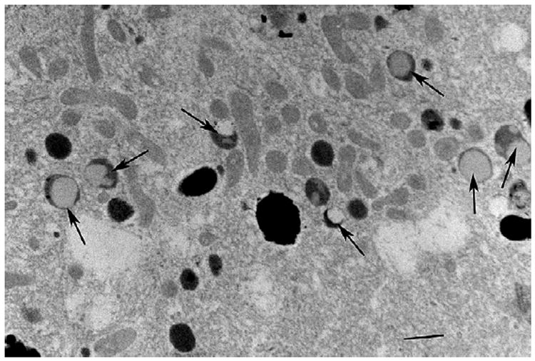
The large phagocytic load that confronts the retinal pigment epithelium (RPE) is thought to play a possible role in the pathogenesis of age related macular degeneration (AMD) that afflicts both humans and monkeys. Our knowledge of how RPE degrades phagosomes and other intra-cellular material by lysosomal action is still rudimentary. In this paper we examine organelles that play a role in this process, melanosome, lysosomes and phagosomes, in the RPE of young and old rhesus monkeys in order to better understand lysosomal autophagy and heterophagy in the RPE and its possible role in AMD. We used electron microscopy to detect and describe the characteristics of melanosomes and lysosome-like organelles in the macular RPE of rhesus monkeys (Macaca mulatta) that were 1, 6, 24, 24, 26 and 35 years of age. The measurements include the number, shape and size of these organelles located in the basal, middle and apical regions of RPE cells. Phaagosomes were also examined but not counted or measured for size or shape because of their rarity. Melanosomes were homogeneously dark with a circular or elliptical shape and decreased in number with age. Smaller melanosomes were more common at the basal side of the RPE. Among the small melanosomes, we found an organelle that was losing melanin in varying degrees; in some cases was nearly devoid of melanin. Because of the melanin loss, we considered this organelle to be a unique type of autophagic melano-lysosome, which we called a Type 1 lysosome. We found another organelle, more canonically lysosomal, which we called a Type 2 lysosome. This organelle was composed of a light matrix containing melanosomes in various stages of degradation. Type 2 lysosomes without melanosomes were rare. Type 2 lysosomes increased while Type 1 decreased in number with age. Phagosomes were rare in both young and old monkeys. They made close contact with Type 2 lysosomes which we considered responsible for their degradation. Melanosomes are being lost from monkey RPE with age. Much of this loss is carried out by two types of lysosomes. One, not defined as unique before, appears to be autophagic in digesting its own melanin; it has been called a Type 1 lysosome. The other, a more canonical lysosome, is both heterophagic in digesting phagosomes and autophagic in digesting local melanosomes; it has been called a Type 2 lysosome. Type 1 lysosomes decrease while type 2 lysosomes increase with age. The loss of melanin is considered to be detrimental to the RPE since it reduces melanin's protective action against light toxicity and oxidative stress. Phagosomes appear to be degraded by membrane contacts with Type 2 lysosomes. The loss of melanin and the buildup of Type 2 lysosomes occur at an earlier age in monkeys than humans implying that a greater vulnerability to senescence accelerates the rate of AMD in monkeys.


Conflict of interest statement

Conflict of interest

None to declare.

Figures

Fig. 1
Fig. 1
Shows melanosomes in monkey RPE. The calibration, lower right, indicates 0.5 microns.
Fig. 2
Fig. 2
Compares the length of the long axis (abscissa) and the number (ordinate) of melanosomes in the basal, middle and apical parts of the RPE cell of the 1, 6, 24 and 35 year old monkeys.
Fig. 3
Fig. 3
Shows examples of Type 1 lysosomes (white arrows). They have a light area in an otherwise densely dark melanosome-like organelle. Other organelles seen are melanosomes (M), mitochondria (mi) and outer segment s (OS). The calibration at the lower left indicates 0.5 microns.
Fig. 4
Fig. 4
Shows a more magnified view of Type 1 lysosomes, several of which have lost virtually all their dark core with only a peripheral rim left. The calibration, middle- left, indicates 0.5 microns.
Fig. 5
Fig. 5
Shows four such melano-lysosomes revealing considerable loss of their dark melanin-like material. The calibration, lower left, indicates 0.1 microns.
Fig. 6
Fig. 6
Compares the length of the long axis (abscissa) and the number (ordinate) of these Type 1 lysosomes in the basal, middle and apical parts of the RPE cell of the 1, 6, 24 and 35 year old monkeys. The 1 year old monkey has many of these organelles.
Fig. 7
Fig. 7
Shows a RPE cell of the 35 year old monkey containing large amounts of Type 2 lysosomes (L2), many not labeled. Most of these organelles have dark inclusion bodies. Two lacking inclusion bodies are marked by black arrows. At the lower right there is a complex of many such Type 2 lysosomes and melanosomes all contained in an amorphous matrix. Mitochondria (mi) are near the basal side and Bruch’s membrane (BM) is below. The calibration, lower right, indicates 1 micron.
Fig. 8
Fig. 8
Shows a Type 2 lysosome in the 35 year old monkey at higher magnification, revealing the many dark inclusion bodies they contain and their slightly irregular perimeters. The calibration, lower left, indicates 0.5 microns.
Fig. 9
Fig. 9
Shows Type 2 lysosomes in the RPE of the 6 year old monkey. The organelles have fewer dark inclusion bodies revealing more of the lighter matrix material and a more regular perimeter. The calibration, lower left, indicates 1 micron.
Fig. 10
Fig. 10
Compares the length of the long axis (abscissa) and the number (ordinate) of Type 2 lysosomes in the basal, middle and apical part of the RPE cell of the 1, 6, 24 and 35 year old monkeys. The 1 year old monkey has none of these organelles.
Fig. 11
Fig. 11
Shows that the cytoplasm of the RPE of the 1year old monkey contains many Type 1 (arrows) but none of the Type 2 lysosomes. The calibration, lower right indicates 0.7 microns.
Fig. 12
Fig. 12
Shows a large elliptical melanosome (lower L) encased by the amorphous matrix of a Type 2 lysosome. Another such lysosome without such a typical melanosome is also present in this section (upper L). The calibration, lower right, indicates 0.4 microns.
Fig. 13
Fig. 13
Shows another example of the engulfing of a typical elongated melanosome by a Type 2 lysosome (L). Another such lysosome (L) but without such a typical melanosome is seen above. The calibration indicates 0.4 microns.
Fig. 14
Fig. 14
Shows a group of Type 2 lysosomes graded for the amount of degradation of the melanosomes they contain. Grade 1 has virtually un-degraded melanosomes; the progressively higher grades show more melanosome degradation to Grade 5 where no large sized melanosomes remain. The calibration, lower left, indicates 0.6 microns.
Fig. 15
Fig. 15
Shows two phagosomes (P) in close contact with Type 2 lysosomes (L). The calibration indicates 0.5 microns.
Fig. 16
Fig. 16
Shows three phagosomes (P) in the RPE of a 24 year old monkey, all in close contact with Type 2 lysosomes (L). There is more degradation of these phagosomes with distance from the apical side of the cell. The calibration, lower left, indicates 0.6 microns.
Fig. 17
Fig. 17
Shows a Type 1 lysosome (large black arrow) that is near but not in close contact with a phagosome (P). Melanosomes (small black arrows) and mitochondria are also seen. The calibration indicates 0.5 microns.

References

    1. Bergmann M, Schutt F, Holz FG, Kopitz J. Inhibition of the ATP driven proton pump in RPE lysosomes by the major lipofuscin fluorophore A2-E may contribute to the pathogenesis of age-related macular degeneration. FASEB J. 2004;18:562–564. - PubMed
    1. Bosch E, Horwitz J, Bok D. Phagocytosis of outer segments by retinal pigment epithelium: phagosome–lysosome interaction. J Histochem Cytochem. 1993;41:253–263. - PubMed
    1. Brandes D, Anton E. Lysosomes in uterine involution: intracytoplasmic degradation of myofilaments and collagen. J Gerontol. 1969;24:55–69. - PubMed
    1. Burke JM, Skumatz MB. Autofluorescent inclusions in long term postconfluent cultures of retinal pigment epithelium. Invest Ophthalmol Vis Sci. 1998;39:1478–1486. - PubMed
    1. Damek-Poprawa M, Diemer T, Vanda S, Lopes VS, Lillo C, Harper DC, Marks MS, Wu Y, Sparrow JR, Rachel RA, Williams DS, Boesze-Battaglia K. Melanoregulin (MREG) modulates lysosome function in pigment epithelial cells. J Cell Biol. 2009;284:10877–10889. - PMC - PubMed

Publication types