. 2012 Jan;194(2):376-94.
doi: 10.1128/JB.06244-11. Epub 2011 Nov 4.

A Rickettsia genome overrun by mobile genetic elements provides insight into the acquisition of genes characteristic of an obligate intracellular lifestyle

Affiliations

A Rickettsia genome overrun by mobile genetic elements provides insight into the acquisition of genes characteristic of an obligate intracellular lifestyle

Joseph J Gillespie et al. J Bacteriol. 2012 Jan.

Abstract

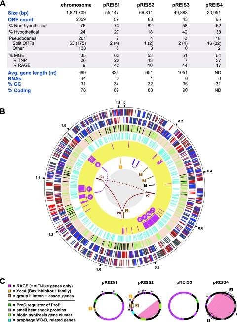
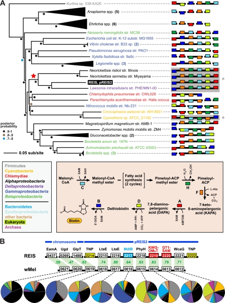
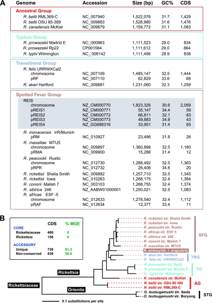
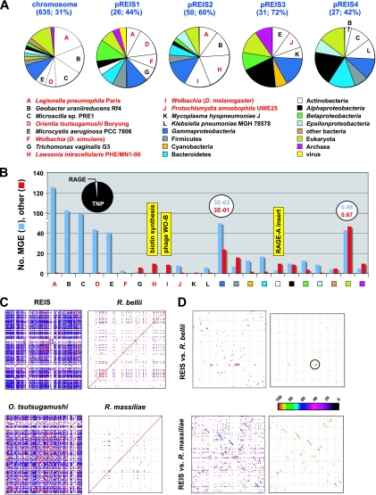
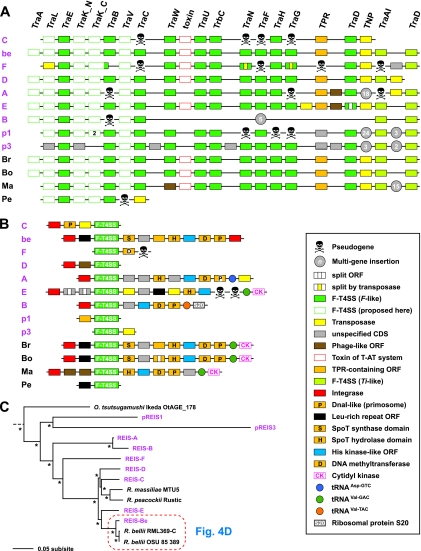
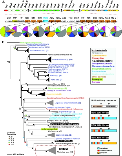
We present the draft genome for the Rickettsia endosymbiont of Ixodes scapularis (REIS), a symbiont of the deer tick vector of Lyme disease in North America. Among Rickettsia species (Alphaproteobacteria: Rickettsiales), REIS has the largest genome sequenced to date (>2 Mb) and contains 2,309 genes across the chromosome and four plasmids (pREIS1 to pREIS4). The most remarkable finding within the REIS genome is the extraordinary proliferation of mobile genetic elements (MGEs), which contributes to a limited synteny with other Rickettsia genomes. In particular, an integrative conjugative element named RAGE (for Rickettsiales amplified genetic element), previously identified in scrub typhus rickettsiae (Orientia tsutsugamushi) genomes, is present on both the REIS chromosome and plasmids. Unlike the pseudogene-laden RAGEs of O. tsutsugamushi, REIS encodes nine conserved RAGEs that include F-like type IV secretion systems similar to that of the tra genes encoded in the Rickettsia bellii and R. massiliae genomes. An unparalleled abundance of encoded transposases (>650) relative to genome size, together with the RAGEs and other MGEs, comprise ~35% of the total genome, making REIS one of the most plastic and repetitive bacterial genomes sequenced to date. We present evidence that conserved rickettsial genes associated with an intracellular lifestyle were acquired via MGEs, especially the RAGE, through a continuum of genomic invasions. Robust phylogeny estimation suggests REIS is ancestral to the virulent spotted fever group of rickettsiae. As REIS is not known to invade vertebrate cells and has no known pathogenic effects on I. scapularis, its genome sequence provides insight on the origin of mechanisms of rickettsial pathogenicity.


Figures

Fig 1
Fig 1
Architecture of the REIS genome. (A) General statistics for the REIS chromosome and four plasmids (pREIS1 to pREIS4). For split genes, total genes are shown with split ORFs in parentheses. (B) Characteristics of the REIS chromosome. The putative origin of replication, positioned at 12 o'clock, was selected based on limited synteny with other SFG genomes (see Fig. S1 in the supplemental material). The outer black circle is a scale with coordinates (in Mb) listed at 200-kb intervals. Six rings inside the scale are the following: plus (1)- and minus (2)-strand genes with core Rickettsia genes colored blue, REIS singletons (not present in other Rickettsiaceae genomes) colored red, and all other genes colored gray; (3) 232 putative pseudogenes colored green (see Table S1 in the supplemental material) and gaps (109 total; see Table S3 in the supplemental material) in the assembly colored black; (4) mobile genetic elements, including transposases, integrases, phage-related ORFs, and other genes typically encoded on plasmids colored turquoise; (5) RAGE genes colored pink, with the location of the seven complete or nearly complete RAGE clusters illustrated (C, Be, F, D, A, E, and B); (6) regions of large-scale duplication and recombination (joined by lines) and regions resulting from plasmid integration (boxed numbers 1 to 3). The ring's six-color scheme is the following (clockwise from the top): purple, duplication of five ORFs and the 6S RNA gene; brown, group II intron and associated genes; black, six-gene insert from pREIS4; burgundy, N-terminal region of rickA (N) joined to the duplicated C-terminal regions (C) (see Fig. S5 in the supplemental material); magenta, four-gene duplication associated with RAGE-A and RAGE-B. (C) Main features of the four REIS plasmids. Duplicated regions within pREIS2 and pREIS4 are depicted with pink shading. Multigene regions (described above) that have been transferred to the chromosome are within dashed boxes. The color scheme depicts genes present on both the chromosome and plasmids (above line) and unique genes of the plasmids discussed in the text.
Fig 2
Fig 2
Two regions of lateral gene transfer (LGT) on pREIS2. (A) LGT of a biotin (bio) operon between the obligate intracellular bacteria REIS, Neorickettsia spp., and Lawsonia intracellularis (noted with a red star at the root). Phylogeny was estimated from the concatenation of six bio genes (bioC, bioH, bioF, bioA, bioD, and bioB; see Table S4 in the supplemental material) from 39 diverse bacteria across seven major taxonomic groups (see the color scheme in the inset at the bottom left; only taxonomic groups above the line are represented in the tree). The schema at the right show the gene order and coding strand for all bio genes per genome, with breaks in black bars denoting noncontiguous genes. The contiguous arrays of all six bio genes in REIS, Neorickettsia spp., Lawsonia intracellularis, and Cyanotheca sp. strain ATCC 51142 are shaded and denoted with an asterisk (see the text for further details). Gene color is described in the inset at the bottom right, which illustrates the recent amendment to the biotin synthesis pathway (75). (B) LGT of a 10-gene region between the prophage WO-B of the Wolbachia endosymbiont of Drosophila melanogaster (wMel) and REIS. EamA, S-adenosylmethionine (SAM) transporter; Ugd, UDP-glucose 6-dehydrogenase; GlpT, glycerol-3-phosphate transporter; LtaE, low-specificity l-threonine aldolase; MdlB, ATP-binding multidrug resistance transporter; PhyH, phytanoyl-CoA dioxygenase; KWG-OMeT, N-terminal KWG repeat domain fused to C-terminal O-methyltransferase (type 2) domain; GT1-SAM, N-terminal glycosyltransferase (type1) domain fused to C-terminal radical SAM domain; WcaG, nucleoside-diphosphate-sugar epimerase. The MdlB ORF is colored blue and is included in the estimated phylogeny shown in Fig. 6. The two domain fusion proteins are colored red and were detected as contiguous ORFs in only one other bacterial genome, Haliangium ochraceum (KWG-OMeT, YP_003265293; GT1-SAM, YP_003265292). The two transposases (TNPs) flanking the pREIS2-carried ORFs are colored yellow. Orthology across REIS and wMel genes is shown with green shading, with percent similarity listed for each comparison. Dashed lines connect each gene with a graphical depiction of the top 100 Blastp subjects using the REIS sequences as queries. The taxonomic color scheme is the same as that in the inset in panel A, with taxa listed from top to bottom (Firmicutes to Archeae) depicted clockwise starting at 12 o'clock on the graphs. Dashed white lines distinguish the Rickettsiales subjects from the remaining Alphaproteobacteria. Additional information pertaining to this region is found in Fig. S2 in the supplemental material.
Fig 3
Fig 3
Rickettsia phylogenomic analysis. (A) Genome statistics for 15 complete Rickettsia genomes and the sequenced plasmid of R. monacensis (7). Statistics were computed from the PATRIC web site (50, 109). Rickettsia spp. are classified according to previous studies (47, 51). The REIS chromosome and plasmids are shaded gray. (B) Phylogeny estimated from 191 concatenated protein alignments (see Materials and Methods for details). STG, scrub typhus group; AG, ancestral group; TG, typhus group; TRG, transitional group; SFG, spotted fever group. The inset illustrates brief results of orthologous group (OG) clustering of 2,309 total predicted REIS ORFs across 16 Rickettsiaceae genomes (see Materials and Methods for details). In green, the percentage of mobile genetic elements per OG category is provided. The complete distribution of OGs across all genomes is provided in Fig. S3 in the supplemental material.
Fig 4
Fig 4
REIS genome plasticity. (A) Taxonomic breakdown of the best non-Rickettsia Blastp hits (n = 769). Homologs from other Rickettsia genomes were either undetected or had a significantly lower E value than the top five non-Rickettsia subjects. For the chromosome and plasmids, the number of total non-Rickettsia hits is provided, followed by the percentage of these sequences per total ORF count. Taxa A to L depict sequences from genomes that are overrepresented on the chromosome and each plasmid, with taxa colored red depicting intracellular species. General taxonomic divisions are colored accordingly, with other bacteria depicting Aquificae (1), Chloroflexi (3), Deinococcus-Thermus (1), Fusobacteria (1), Spirochaetes (1), and Tenericutes (2). (B) Number of molecular genetic elements (MGEs) and other CDS per taxon or taxonomic category listed in panel A. For taxa A to E, the 414 total MGEs are further divided into transposases and related elements (TNPs, 405) and components of the RAGE (7) as illustrated in the pie chart. Average E values are shown for both the MGEs and other CDS for the Gammaproteobacteria and Eukarya hits, illustrating the low average significance of the latter. Three regions of lateral gene transfer in the REIS genome are mapped to their respective taxon as illustrated in yellow (see the text for details). (C) Plots of sequence repeat density in four select Rickettsiales genomes. Each dot represents a repeat (stretches of similar sequences located in multiple regions of a genome), with blue depicting direct repeats and red depicting reverse repeats. (D) Genome synteny and comparative analysis of repeat density for REIS versus R. bellii strain RML369-C (top) and REIS versus R. massiliae strain MTU5 (bottom). The color scheme for plots at the left is the same as that for panel C, and the color scheme at the right follows the color spectrum depicting percent sequence identity. The high similarity between the RAGE of R. bellii and REIS RAGE-Be is encircled, with the monophyly of these two elements relative to the remaining RAGEs being shown in Fig. 5C.
Fig 5
Fig 5
Characteristics of the RAGE. (A and B) Schema of gene organization within nine complete (or nearly complete) RAGEs in the REIS genome (pink) and their comparison to the single-copy RAGEs encoded in four other Rickettsia genomes: Br, R. bellii strain RML369-C; Bo, R. bellii strain OSU 85-389; Ma, R. massiliae strain MTU5; Pe, R. peacockii strain Rustic. The genome coordinates of the REIS RAGEs are illustrated in Fig. 1B. Gene color and symbols are described in the inset. (A) Illustration of the RAGE-encoded F-like type IV secretion system (F-T4SS) genes. (B) Illustration of the regions flanking the RAGE F-T4SS genes, including several proposed tRNA insertion sites. (C) Phylogeny estimation of the RAGE genes illustrated in panel A. Nineteen protein families were included in the analysis (excluding TraA pilins and the conserved transposase flanked by TraDF and TraAITi), with pseudogenes not included. Branch support (posterior probabilities) is from Bayesian analysis (see the text for details). Phylogeny estimates of the individual RAGE genes are provided in Fig. S10 in the supplemental material.
Fig 6
Fig 6
Eighteen-gene insert piggybacking on RAGE-A. (A) The schema at the top depicts RAGE-A, and the color scheme and symbols are explained in Fig. 5. Red dashed lines illustrate an 18-gene insert between traDF (F plasmid-like) and traAITi (Ti plasmid-like). ORFs colored orange have the closest homology to components typical of aminoglycoside antibiotic biosynthesis gene clusters: IstN, SAM-methyltransferase; IstM, NDP-d-glucosaminyltransferase; BtrR, l-glutamine:deoxy-scyllo-inosose (DOI) aminotransferase; AprU, apramycin kinase; HemL, glutamate-1-semialdehyde aminotransferase; IstC, 2-deoxy-scyllo-inosose synthase; Tdh, l-threonine 3-dehydrogenase; Sis6, sugar-alcohol dehydrogenase; TbmB, l-glutamine:DOI aminotransferase; PIG-L, GlcNAc-PI de-N-acetylase. Dashed lines connect each gene with a graphical depiction of the top 100 Blastp subjects using the REIS sequences as queries. The taxonomic color scheme follows the inset at the bottom, with taxa listed from top to bottom (Actinobacteria to Archeae) depicted clockwise starting at 12 o'clock on the graphs. Dashed white lines distinguish the Rickettsiales subjects from the remaining Alphaproteobacteria. The top five Blastp hits for each of these ORFs are listed in Table S10 in the supplemental material. The MdlB ORF is colored blue and included in the estimated phylogeny in panel B. An associated transposase (TNP) is colored yellow. Additional ORFs are GlpT, glycerol-3-phosphate transporter (see Fig. 2B); HP, hypothetical protein; ABC, uncharacterized ABC transporter (COG4178); Tlc, ADP/ATP translocase (rickettsial type 2 Tlc); and LuxR, HTH transcriptional activator. Additional information pertaining to ABC, Tlc, and LuxR is provided in Fig. S11 in the supplemental material. (B) Estimated phylogeny of an uncharacterized multidrug resistance transporter, MdlB (EC 3.6.3.44). A gray star depicts the monophyly of mdlB genes within diverse intracellular bacterial species, with these lineages colored gray (except for the eukaryote Hydra magnipapilata). The three REIS ORFs are boxed, with a red dashed box enclosing the ortholog conserved in all Rickettsia genomes. The predicted membrane association of all three REIS orthologs is shown at the right, with TMS regions depicted using TMHMM v.2.0 (70).

References

    1. Adelson ME, et al. 2004. Prevalence of Borrelia burgdorferi, Bartonella spp., Babesia microti, and Anaplasma phagocytophila in Ixodes scapularis ticks collected in Northern New Jersey. J. Clin. Microbiol. 42: 2799–2801 - PMC - PubMed
    1. Andersson SG, Stothard DR, Fuerst P, Kurland CG. 1999. Molecular phylogeny and rearrangement of rRNA genes in Rickettsia species. Mol. Biol. Evol. 16: 987–995 - PubMed
    1. Andersson SG, et al. 1998. The genome sequence of Rickettsia prowazekii and the origin of mitochondria. Nature 396: 133–140 - PubMed
    1. Audia JP, Winkler HH. 2006. Study of the five Rickettsia prowazekii proteins annotated as ATP/ADP translocases (Tlc): only Tlc1 transports ATP/ADP, while Tlc4 and Tlc5 transport other ribonucleotides. J. Bacteriol. 188: 6261–6268 - PMC - PubMed
    1. Aziz RK, et al. 2008. The RAST server: rapid annotations using subsystems technology. BMC Genomics 9: 75. - PMC - PubMed

Publication types

LinkOut - more resources