

MYC gene amplification is often acquired in lethal distant breast cancer metastases of unamplified primary tumors
- PMID: 22056952
- PMCID: PMC3276715
- DOI: 10.1038/modpathol.2011.171
MYC gene amplification is often acquired in lethal distant breast cancer metastases of unamplified primary tumors
Abstract
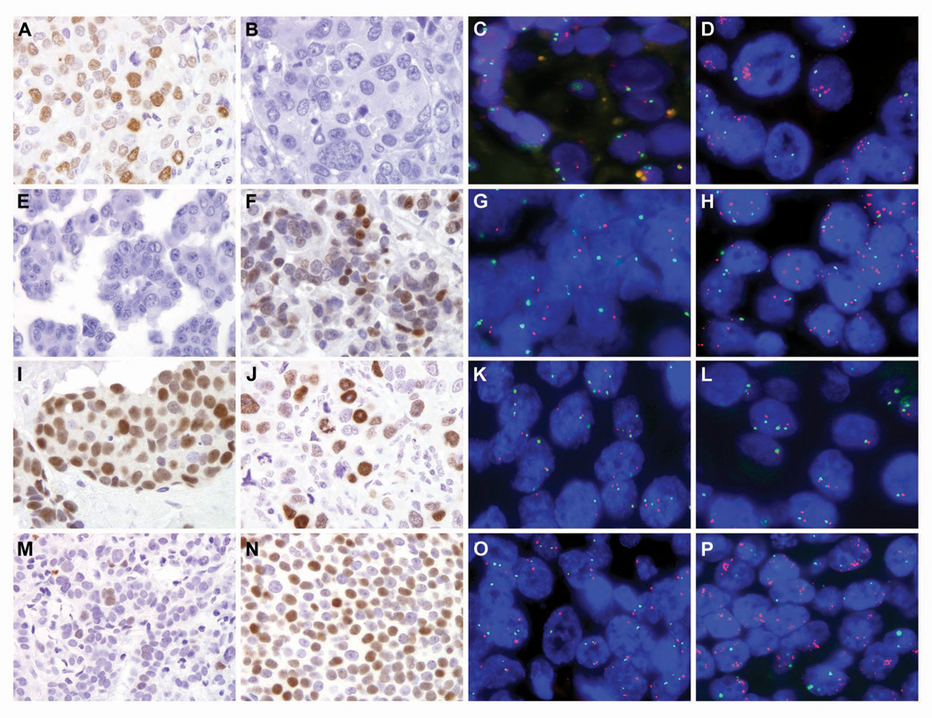
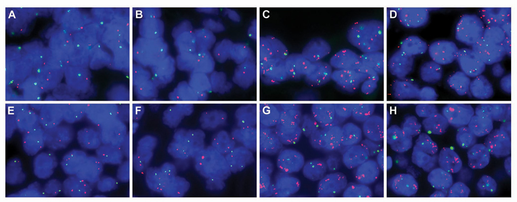
In breast cancer, amplification of MYC is consistently observed in aggressive forms of disease and correlates with poor prognosis and distant metastases. However, to date, a systematic analysis of MYC amplification in metastatic breast cancers has not been reported. Specifically, whether the MYC amplification status may change in metastases in comparison to the corresponding primary breast tumor, and potential variability among different metastases within the same patient have also not been assessed. We generated single patient tissue microarrays consisting of both primary breast carcinomas and multiple matched systemic metastases from 15 patients through our previously described rapid autopsy program. In total, the 15 tissue microarrays contained 145 primary tumor spots and 778 spots derived from 180 different metastases. In addition, two separate tissue microarrays were constructed composed of 10 matched primary breast cancers and corresponding solitary metastases sampled not at autopsy but rather in routine surgical resections. These two tissue microarrays totaled 50 primary tumor spots and 86 metastatic tumor spots. For each case, hormone receptor status, HER2/neu, EGFR and CK5/6 expression were assessed, and the cases were characterized as luminal, basal-like or HER2 based on published criteria. Both fluorescence in situ hybridization and immunohistochemistry for MYC was performed on all cases. Of the 25 cases, 24 were evaluable. While 4 of 24 primary tumors (16%) demonstrated MYC amplification, an additional 6 (25% of total evaluable cases) acquired MYC amplification in their systemic metastases. Of note, there was remarkably little heterogeneity in MYC copy number among different metastases from the same patient. MYC immunoreactivity was increased in metastases relative to matched primaries in the surgical cohort, although there was no perfect correlation with MYC amplification. In conclusion, amplification of MYC is a frequent event in breast cancer, but occurs more frequently as a diffuse, acquired event in metastatic disease than in the corresponding primary. These observations underscore the importance of MYC in breast cancer progression/metastasis, as well as its relevance as a potential therapeutic target in otherwise incurable metastatic disease.
Figures
References
-
- Tirkkonen M, Tanner M, Karhu R, et al. Molecular cytogenetics of primary breast cancer by CGH. Genes Chromosomes Cancer. 1998;21:177–184. - PubMed
-
- Courjal F, Theillet C. Comparative genomic hybridization analysis of breast tumors with predetermined profiles of DNA amplification. Cancer Res. 1997;57:4368–4377. - PubMed
-
- Nishizaki T, DeVries S, Chew K, et al. Genetic alterations in primary breast cancers and their metastases: direct comparison using modified comparative genomic hybridization. Genes Chromosomes Cancer. 1997;19:267–272. - PubMed
Publication types
MeSH terms
Substances
Grants and funding
LinkOut - more resources
Full Text Sources
Medical
Research Materials
Miscellaneous
